Cadmium »
PDB 1a4k-1dmf »
1aaz »
Cadmium in PDB 1aaz: The Structure of Oxidized Bacteriophage T4 Glutaredoxin (Thioredoxin)
Protein crystallography data
The structure of The Structure of Oxidized Bacteriophage T4 Glutaredoxin (Thioredoxin), PDB code: 1aaz
was solved by
H.Eklund,
M.Ingelman,
B.-O.Soderberg,
T.Uhlin,
P.Nordlund,
M.Nikkola,
U.Sonnerstam,
T.Joelson,
K.Petratos,
with X-Ray Crystallography technique. A brief refinement statistics is given in the table below:
| Resolution Low / High (Å) | N/A / 2.00 |
| Space group | P 1 21 1 |
| Cell size a, b, c (Å), α, β, γ (°) | 54.100, 45.900, 40.800, 90.00, 99.40, 90.00 |
| R / Rfree (%) | 21 / n/a |
Cadmium Binding Sites:
The binding sites of Cadmium atom in the The Structure of Oxidized Bacteriophage T4 Glutaredoxin (Thioredoxin)
(pdb code 1aaz). This binding sites where shown within
5.0 Angstroms radius around Cadmium atom.
In total 2 binding sites of Cadmium where determined in the The Structure of Oxidized Bacteriophage T4 Glutaredoxin (Thioredoxin), PDB code: 1aaz:
Jump to Cadmium binding site number: 1; 2;
In total 2 binding sites of Cadmium where determined in the The Structure of Oxidized Bacteriophage T4 Glutaredoxin (Thioredoxin), PDB code: 1aaz:
Jump to Cadmium binding site number: 1; 2;
Cadmium binding site 1 out of 2 in 1aaz
Go back to
Cadmium binding site 1 out
of 2 in the The Structure of Oxidized Bacteriophage T4 Glutaredoxin (Thioredoxin)
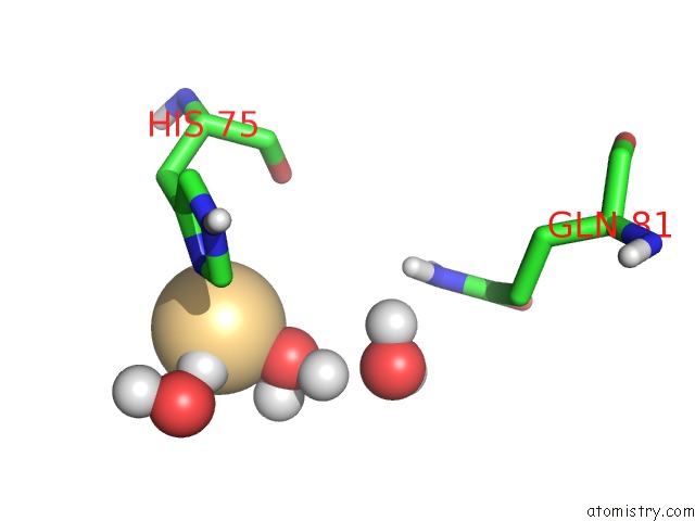
Mono view

Stereo pair view
Mono view
Stereo pair view
A full contact list of Cadmium with other atoms in the Cd binding
site number 1 of The Structure of Oxidized Bacteriophage T4 Glutaredoxin (Thioredoxin) within 5.0Å range:
|
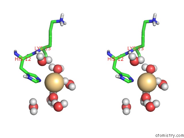
Cadmium binding site 2 out of 2 in 1aaz
Go back to
Cadmium binding site 2 out
of 2 in the The Structure of Oxidized Bacteriophage T4 Glutaredoxin (Thioredoxin)
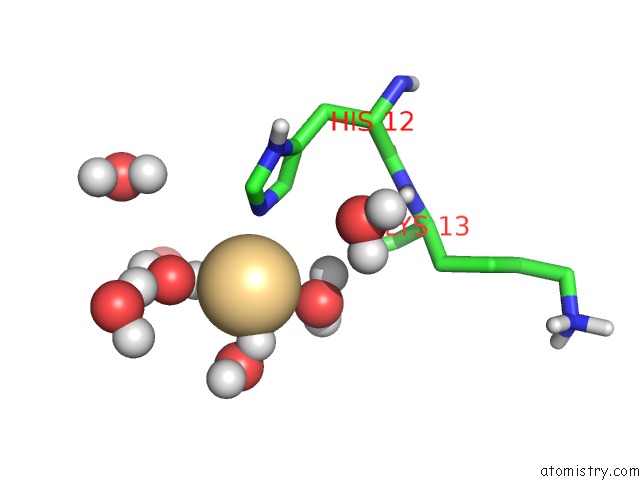
Mono view
Stereo pair view
Mono view
Stereo pair view
A full contact list of Cadmium with other atoms in the Cd binding
site number 2 of The Structure of Oxidized Bacteriophage T4 Glutaredoxin (Thioredoxin) within 5.0Å range:
|
Reference:
H.Eklund,
M.Ingelman,
B.O.Soderberg,
T.Uhlin,
P.Nordlund,
M.Nikkola,
U.Sonnerstam,
T.Joelson,
K.Petratos.
Structure of Oxidized Bacteriophage T4 Glutaredoxin (Thioredoxin). Refinement of Native and Mutant Proteins. J.Mol.Biol. V. 228 596 1992.
ISSN: ISSN 0022-2836
PubMed: 1453466
DOI: 10.1016/0022-2836(92)90844-A
Page generated: Thu Jul 10 10:28:14 2025
ISSN: ISSN 0022-2836
PubMed: 1453466
DOI: 10.1016/0022-2836(92)90844-A
Last articles
Cl in 7W5TCl in 7W5V
Cl in 7W5S
Cl in 7W06
Cl in 7VXG
Cl in 7VZY
Cl in 7VZZ
Cl in 7VYQ
Cl in 7VZN
Cl in 7VZQ