Cadmium »
PDB 1dpe-1gwg »
1eu1 »
Cadmium in PDB 1eu1: The Crystal Structure of Rhodobacter Sphaeroides Dimethylsulfoxide Reductase Reveals Two Distinct Molybdenum Coordination Environments.
Protein crystallography data
The structure of The Crystal Structure of Rhodobacter Sphaeroides Dimethylsulfoxide Reductase Reveals Two Distinct Molybdenum Coordination Environments., PDB code: 1eu1
was solved by
H.K.Li,
K.Temple,
K.V.Rajagopalan,
H.Schindelin,
with X-Ray Crystallography technique. A brief refinement statistics is given in the table below:
| Resolution Low / High (Å) | 10.00 / 1.30 |
| Space group | P 21 21 2 |
| Cell size a, b, c (Å), α, β, γ (°) | 102.176, 141.718, 59.812, 90.00, 90.00, 90.00 |
| R / Rfree (%) | 12.1 / 14.5 |
Other elements in 1eu1:
The structure of The Crystal Structure of Rhodobacter Sphaeroides Dimethylsulfoxide Reductase Reveals Two Distinct Molybdenum Coordination Environments. also contains other interesting chemical elements:
| Molybdenum | (Mo) | 2 atoms |
Cadmium Binding Sites:
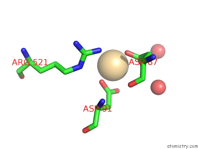
The binding sites of Cadmium atom in the The Crystal Structure of Rhodobacter Sphaeroides Dimethylsulfoxide Reductase Reveals Two Distinct Molybdenum Coordination Environments.
(pdb code 1eu1). This binding sites where shown within
5.0 Angstroms radius around Cadmium atom.
In total only one binding site of Cadmium was determined in the The Crystal Structure of Rhodobacter Sphaeroides Dimethylsulfoxide Reductase Reveals Two Distinct Molybdenum Coordination Environments., PDB code: 1eu1:
In total only one binding site of Cadmium was determined in the The Crystal Structure of Rhodobacter Sphaeroides Dimethylsulfoxide Reductase Reveals Two Distinct Molybdenum Coordination Environments., PDB code: 1eu1:
Cadmium binding site 1 out of 1 in 1eu1
Go back to
Cadmium binding site 1 out
of 1 in the The Crystal Structure of Rhodobacter Sphaeroides Dimethylsulfoxide Reductase Reveals Two Distinct Molybdenum Coordination Environments.
Mono view
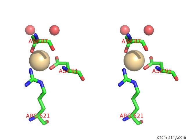
Stereo pair view
Mono view
Stereo pair view
A full contact list of Cadmium with other atoms in the Cd binding
site number 1 of The Crystal Structure of Rhodobacter Sphaeroides Dimethylsulfoxide Reductase Reveals Two Distinct Molybdenum Coordination Environments. within 5.0Å range:
|
Reference:
H.K.Li,
K.Temple,
K.V.Rajagopalan,
H.Schindelin.
The 1.3 A Crystal Structure of Rhodobacter Sphaeroides Dimethylsulfoxide Reductase Reveals Two Distinct Molybdenum Coordination Environments J.Am.Chem.Soc. V. 122 7673 2000.
ISSN: ISSN 0002-7863
DOI: 10.1021/JA000643E
Page generated: Fri Jul 19 13:15:58 2024
ISSN: ISSN 0002-7863
DOI: 10.1021/JA000643E
Last articles
Zn in 9MJ5Zn in 9HNW
Zn in 9G0L
Zn in 9FNE
Zn in 9DZN
Zn in 9E0I
Zn in 9D32
Zn in 9DAK
Zn in 8ZXC
Zn in 8ZUF