Cadmium »
PDB 1gyn-1jre »
1hb6 »
Cadmium in PDB 1hb6: Structure of Bovine Acyl-Coa Binding Protein in Orthorhombic Crystal Form
Protein crystallography data
The structure of Structure of Bovine Acyl-Coa Binding Protein in Orthorhombic Crystal Form, PDB code: 1hb6
was solved by
J.Y.Zou,
G.J.Kleywegt,
T.Bergfors,
J.Knudsen,
T.A.Jones,
with X-Ray Crystallography technique. A brief refinement statistics is given in the table below:
| Resolution Low / High (Å) | 19.14 / 2.0 |
| Space group | P 21 21 21 |
| Cell size a, b, c (Å), α, β, γ (°) | 26.050, 54.780, 65.830, 90.00, 90.00, 90.00 |
| R / Rfree (%) | 20 / 22.6 |
Cadmium Binding Sites:
The binding sites of Cadmium atom in the Structure of Bovine Acyl-Coa Binding Protein in Orthorhombic Crystal Form
(pdb code 1hb6). This binding sites where shown within
5.0 Angstroms radius around Cadmium atom.
In total only one binding site of Cadmium was determined in the Structure of Bovine Acyl-Coa Binding Protein in Orthorhombic Crystal Form, PDB code: 1hb6:
In total only one binding site of Cadmium was determined in the Structure of Bovine Acyl-Coa Binding Protein in Orthorhombic Crystal Form, PDB code: 1hb6:
Cadmium binding site 1 out of 1 in 1hb6
Go back to
Cadmium binding site 1 out
of 1 in the Structure of Bovine Acyl-Coa Binding Protein in Orthorhombic Crystal Form
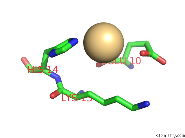
Mono view
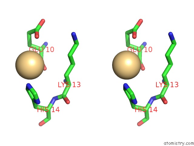
Stereo pair view
Mono view
Stereo pair view
A full contact list of Cadmium with other atoms in the Cd binding
site number 1 of Structure of Bovine Acyl-Coa Binding Protein in Orthorhombic Crystal Form within 5.0Å range:
|
Reference:
D.M.F.Van Aalten,
K.G.Milne,
J.Y.Zou,
G.J.Kleywegt,
T.Bergfors,
M.A.J.Ferguson,
J.Knudsen,
T.A.Jones.
Binding Site Differences Revealed By Crystal Structures of Plasmodium Falciparum and Bovine Acyl-Coa Binding Protein J.Mol.Biol. V. 309 181 2001.
ISSN: ISSN 0022-2836
PubMed: 11491287
DOI: 10.1006/JMBI.2001.4749
Page generated: Thu Jul 10 10:46:26 2025
ISSN: ISSN 0022-2836
PubMed: 11491287
DOI: 10.1006/JMBI.2001.4749
Last articles
F in 7MT6F in 7MSA
F in 7MTY
F in 7MT5
F in 7MSO
F in 7MSD
F in 7MT4
F in 7MSB
F in 7MSK
F in 7MS5