Cadmium »
PDB 1jv4-1mwr »
1m90 »
Cadmium in PDB 1m90: Co-Crystal Structure of Cca-Phe-Caproic Acid-Biotin and Sparsomycin Bound to the 50S Ribosomal Subunit
Protein crystallography data
The structure of Co-Crystal Structure of Cca-Phe-Caproic Acid-Biotin and Sparsomycin Bound to the 50S Ribosomal Subunit, PDB code: 1m90
was solved by
J.L.Hansen,
T.M.Schmeing,
P.B.Moore,
T.A.Steitz,
with X-Ray Crystallography technique. A brief refinement statistics is given in the table below:
| Resolution Low / High (Å) | 20.00 / 2.80 |
| Space group | C 2 2 21 |
| Cell size a, b, c (Å), α, β, γ (°) | 211.661, 299.773, 573.684, 90.00, 90.00, 90.00 |
| R / Rfree (%) | 18.1 / 22.2 |
Other elements in 1m90:
The structure of Co-Crystal Structure of Cca-Phe-Caproic Acid-Biotin and Sparsomycin Bound to the 50S Ribosomal Subunit also contains other interesting chemical elements:
| Magnesium | (Mg) | 119 atoms |
| Potassium | (K) | 2 atoms |
| Chlorine | (Cl) | 22 atoms |
| Sodium | (Na) | 86 atoms |
Cadmium Binding Sites:
The binding sites of Cadmium atom in the Co-Crystal Structure of Cca-Phe-Caproic Acid-Biotin and Sparsomycin Bound to the 50S Ribosomal Subunit
(pdb code 1m90). This binding sites where shown within
5.0 Angstroms radius around Cadmium atom.
In total 5 binding sites of Cadmium where determined in the Co-Crystal Structure of Cca-Phe-Caproic Acid-Biotin and Sparsomycin Bound to the 50S Ribosomal Subunit, PDB code: 1m90:
Jump to Cadmium binding site number: 1; 2; 3; 4; 5;
In total 5 binding sites of Cadmium where determined in the Co-Crystal Structure of Cca-Phe-Caproic Acid-Biotin and Sparsomycin Bound to the 50S Ribosomal Subunit, PDB code: 1m90:
Jump to Cadmium binding site number: 1; 2; 3; 4; 5;
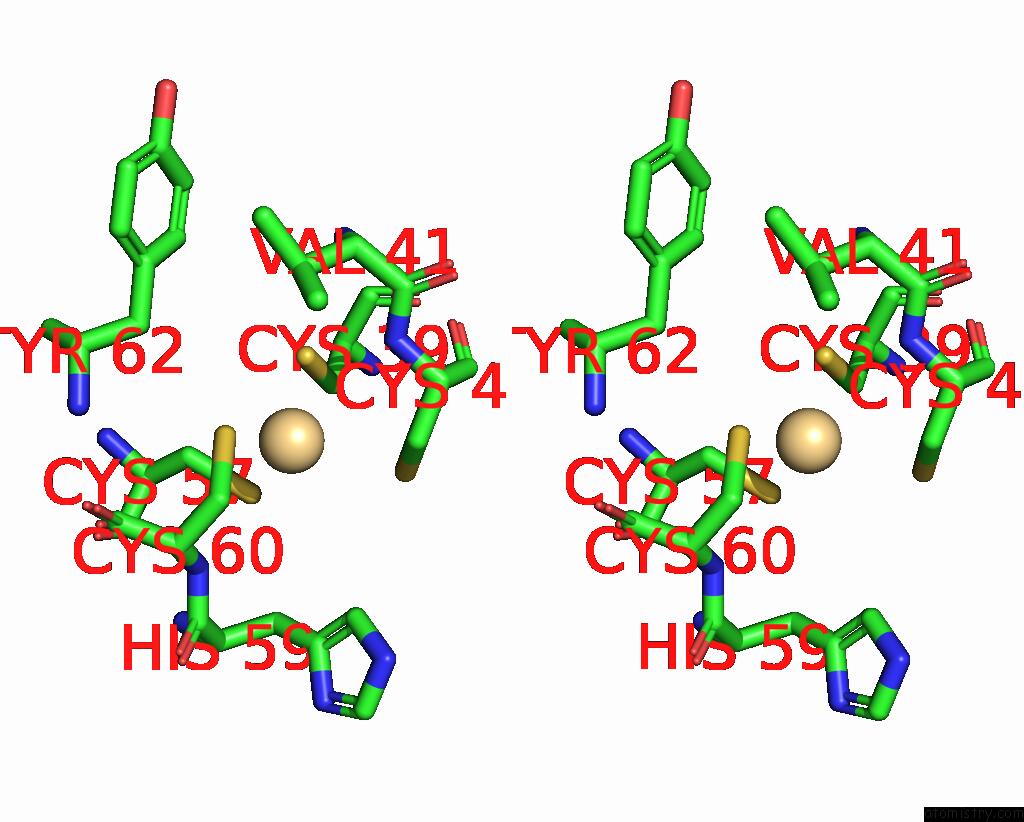
Cadmium binding site 1 out of 5 in 1m90
Go back to
Cadmium binding site 1 out
of 5 in the Co-Crystal Structure of Cca-Phe-Caproic Acid-Biotin and Sparsomycin Bound to the 50S Ribosomal Subunit
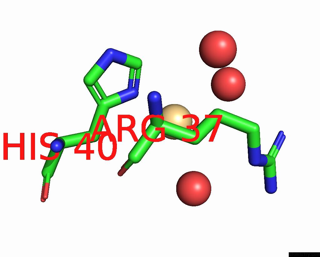
Mono view
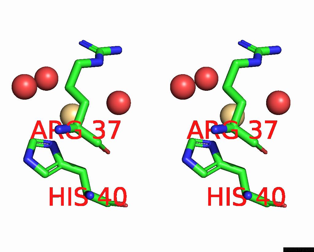
Stereo pair view
Mono view
Stereo pair view
A full contact list of Cadmium with other atoms in the Cd binding
site number 1 of Co-Crystal Structure of Cca-Phe-Caproic Acid-Biotin and Sparsomycin Bound to the 50S Ribosomal Subunit within 5.0Å range:
|
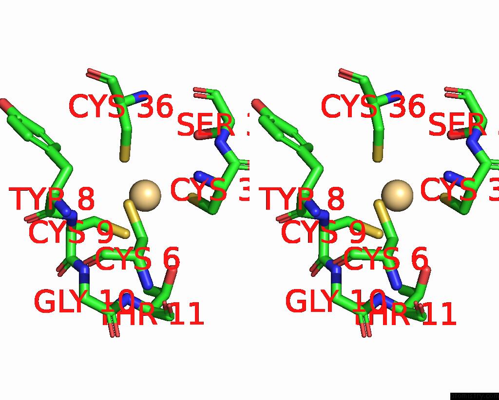
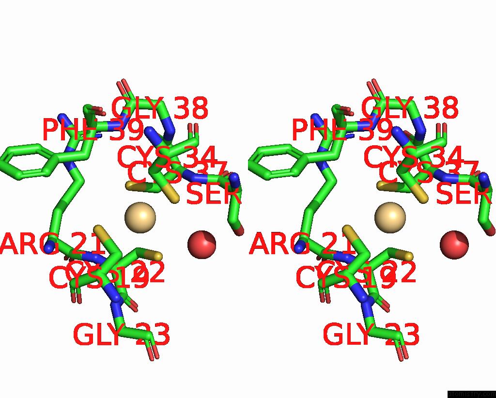
Cadmium binding site 2 out of 5 in 1m90
Go back to
Cadmium binding site 2 out
of 5 in the Co-Crystal Structure of Cca-Phe-Caproic Acid-Biotin and Sparsomycin Bound to the 50S Ribosomal Subunit

Mono view
Stereo pair view
Mono view
Stereo pair view
A full contact list of Cadmium with other atoms in the Cd binding
site number 2 of Co-Crystal Structure of Cca-Phe-Caproic Acid-Biotin and Sparsomycin Bound to the 50S Ribosomal Subunit within 5.0Å range:
|
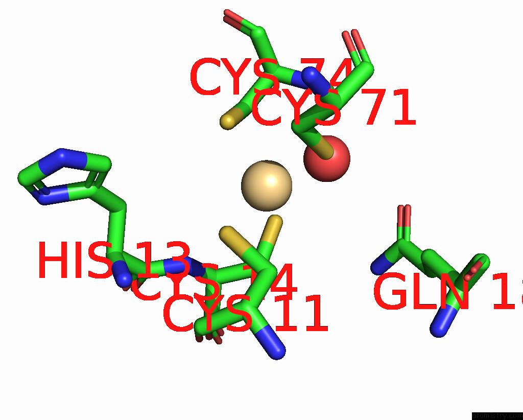
Cadmium binding site 3 out of 5 in 1m90
Go back to
Cadmium binding site 3 out
of 5 in the Co-Crystal Structure of Cca-Phe-Caproic Acid-Biotin and Sparsomycin Bound to the 50S Ribosomal Subunit
Mono view
Stereo pair view
Mono view
Stereo pair view
A full contact list of Cadmium with other atoms in the Cd binding
site number 3 of Co-Crystal Structure of Cca-Phe-Caproic Acid-Biotin and Sparsomycin Bound to the 50S Ribosomal Subunit within 5.0Å range:
|
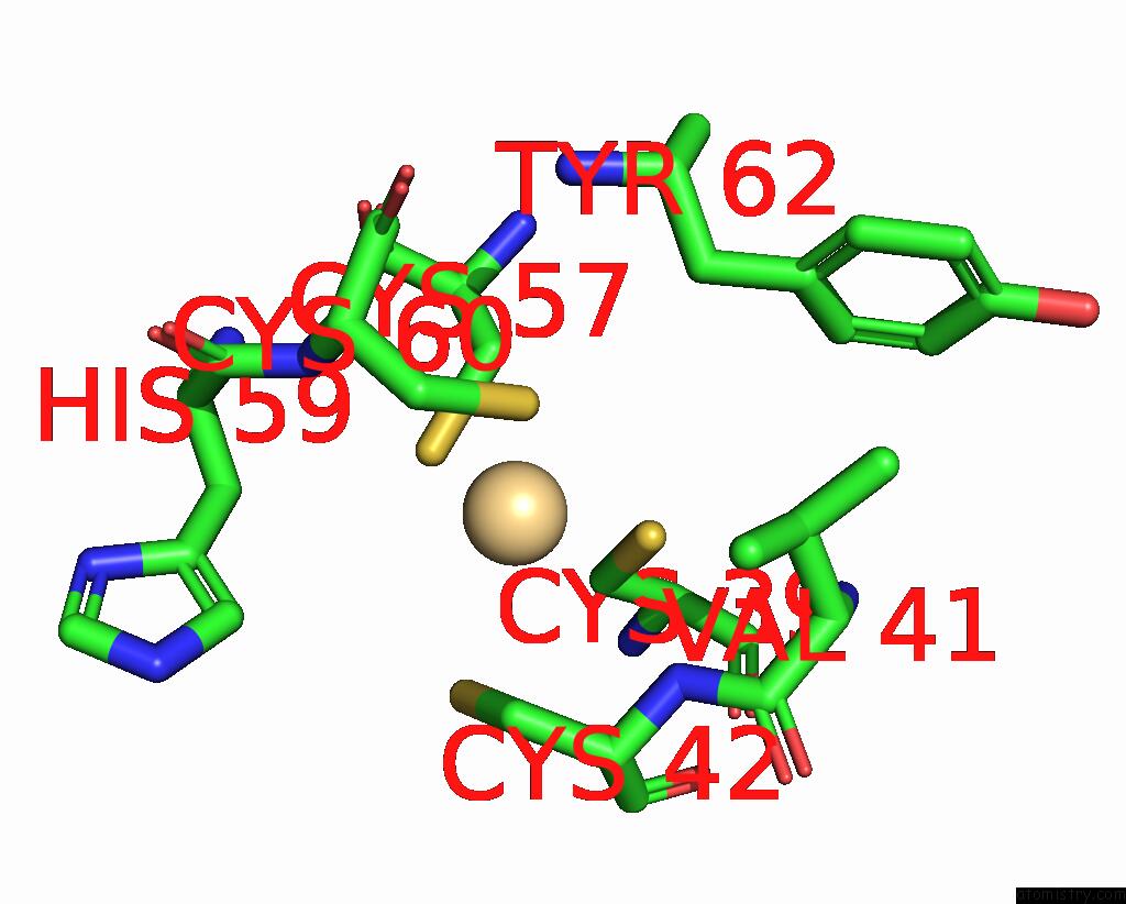
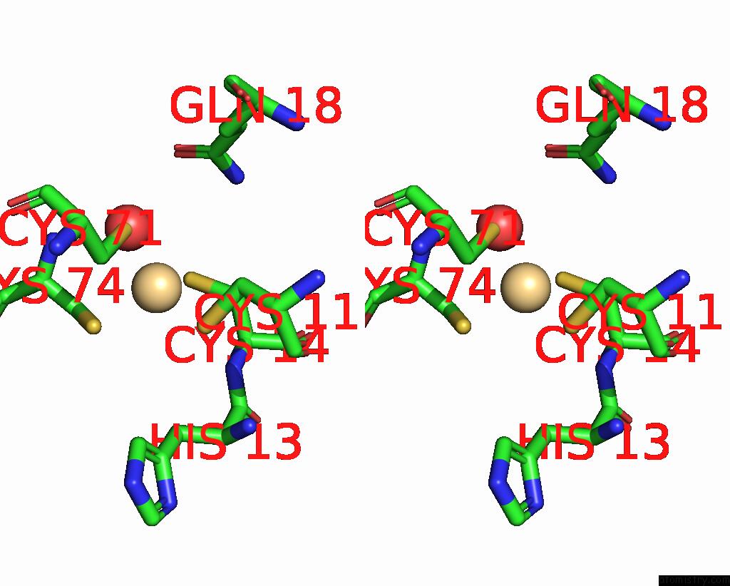
Cadmium binding site 4 out of 5 in 1m90
Go back to
Cadmium binding site 4 out
of 5 in the Co-Crystal Structure of Cca-Phe-Caproic Acid-Biotin and Sparsomycin Bound to the 50S Ribosomal Subunit

Mono view
Stereo pair view
Mono view
Stereo pair view
A full contact list of Cadmium with other atoms in the Cd binding
site number 4 of Co-Crystal Structure of Cca-Phe-Caproic Acid-Biotin and Sparsomycin Bound to the 50S Ribosomal Subunit within 5.0Å range:
|
Cadmium binding site 5 out of 5 in 1m90
Go back to
Cadmium binding site 5 out
of 5 in the Co-Crystal Structure of Cca-Phe-Caproic Acid-Biotin and Sparsomycin Bound to the 50S Ribosomal Subunit
Mono view
Stereo pair view
Mono view
Stereo pair view
A full contact list of Cadmium with other atoms in the Cd binding
site number 5 of Co-Crystal Structure of Cca-Phe-Caproic Acid-Biotin and Sparsomycin Bound to the 50S Ribosomal Subunit within 5.0Å range:
|
Reference:
J.L.Hansen,
T.M.Schmeing,
P.B.Moore,
T.A.Steitz.
Structural Insights Into Peptide Bond Formation. Proc.Natl.Acad.Sci.Usa V. 99 11670 2002.
ISSN: ISSN 0027-8424
PubMed: 12185246
DOI: 10.1073/PNAS.172404099
Page generated: Fri Jul 19 13:51:54 2024
ISSN: ISSN 0027-8424
PubMed: 12185246
DOI: 10.1073/PNAS.172404099
Last articles
Zn in 9MJ5Zn in 9HNW
Zn in 9G0L
Zn in 9FNE
Zn in 9DZN
Zn in 9E0I
Zn in 9D32
Zn in 9DAK
Zn in 8ZXC
Zn in 8ZUF