Cadmium in PDB 1mhu: The Three-Dimensional Structure of Human [113CD7] Metallothionein-2 in Solution Determined By Nuclear Magnetic Resonance Spectroscopy
Cadmium Binding Sites:
The binding sites of Cadmium atom in the The Three-Dimensional Structure of Human [113CD7] Metallothionein-2 in Solution Determined By Nuclear Magnetic Resonance Spectroscopy
(pdb code 1mhu). This binding sites where shown within
5.0 Angstroms radius around Cadmium atom.
In total 4 binding sites of Cadmium where determined in the
The Three-Dimensional Structure of Human [113CD7] Metallothionein-2 in Solution Determined By Nuclear Magnetic Resonance Spectroscopy, PDB code: 1mhu:
Jump to Cadmium binding site number:
1;
2;
3;
4;
Cadmium binding site 1 out
of 4 in 1mhu
Go back to
Cadmium Binding Sites List in 1mhu
Cadmium binding site 1 out
of 4 in the The Three-Dimensional Structure of Human [113CD7] Metallothionein-2 in Solution Determined By Nuclear Magnetic Resonance Spectroscopy
Mono view
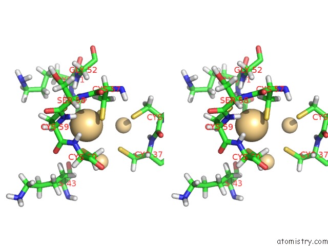
Stereo pair view
|
A full contact list of Cadmium with other atoms in the Cd binding
site number 1 of The Three-Dimensional Structure of Human [113CD7] Metallothionein-2 in Solution Determined By Nuclear Magnetic Resonance Spectroscopy within 5.0Å range:
probe
|
atom
|
residue
|
distance (Å)
|
B
|
Occ
|
A:Cd101
b:1.0
occ:1.00
|
SG
|
A:CYS60
|
2.5
|
1.4
|
1.0
|
SG
|
A:CYS57
|
2.6
|
1.2
|
1.0
|
SG
|
A:CYS59
|
2.6
|
1.7
|
1.0
|
SG
|
A:CYS50
|
2.7
|
0.6
|
1.0
|
H
|
A:CYS59
|
2.7
|
1.1
|
1.0
|
HB2
|
A:CYS57
|
3.0
|
0.8
|
1.0
|
HB2
|
A:CYS60
|
3.0
|
1.2
|
1.0
|
H
|
A:CYS60
|
3.1
|
1.4
|
1.0
|
N
|
A:CYS60
|
3.1
|
1.4
|
1.0
|
HA
|
A:CYS50
|
3.1
|
0.6
|
1.0
|
HB2
|
A:CYS50
|
3.2
|
1.2
|
1.0
|
CB
|
A:CYS60
|
3.2
|
1.4
|
1.0
|
CB
|
A:CYS57
|
3.3
|
0.9
|
1.0
|
C
|
A:CYS59
|
3.3
|
1.3
|
1.0
|
CB
|
A:CYS50
|
3.4
|
0.8
|
1.0
|
CB
|
A:CYS59
|
3.4
|
1.0
|
1.0
|
HB2
|
A:CYS59
|
3.4
|
1.2
|
1.0
|
N
|
A:CYS59
|
3.5
|
1.3
|
1.0
|
CA
|
A:CYS59
|
3.5
|
1.2
|
1.0
|
HB3
|
A:CYS57
|
3.6
|
0.8
|
1.0
|
H
|
A:SER58
|
3.7
|
1.7
|
1.0
|
CA
|
A:CYS60
|
3.8
|
1.6
|
1.0
|
CA
|
A:CYS50
|
3.8
|
0.6
|
1.0
|
O
|
A:CYS59
|
3.9
|
1.4
|
1.0
|
H
|
A:GLY52
|
4.1
|
3.6
|
1.0
|
SG
|
A:CYS37
|
4.1
|
1.7
|
1.0
|
HB3
|
A:CYS60
|
4.2
|
1.6
|
1.0
|
H
|
A:LYS51
|
4.3
|
1.7
|
1.0
|
CD
|
A:CD107
|
4.3
|
0.9
|
1.0
|
CD
|
A:CD106
|
4.3
|
1.4
|
1.0
|
N
|
A:SER58
|
4.4
|
1.6
|
1.0
|
HB3
|
A:CYS50
|
4.4
|
1.2
|
1.0
|
HB3
|
A:CYS59
|
4.4
|
1.6
|
1.0
|
HA
|
A:CYS60
|
4.5
|
1.9
|
1.0
|
SG
|
A:CYS36
|
4.6
|
3.2
|
1.0
|
HA3
|
A:GLY52
|
4.6
|
5.6
|
1.0
|
HA
|
A:CYS59
|
4.6
|
1.4
|
1.0
|
CA
|
A:CYS57
|
4.6
|
1.2
|
1.0
|
C
|
A:SER58
|
4.7
|
1.7
|
1.0
|
C
|
A:CYS50
|
4.7
|
0.8
|
1.0
|
N
|
A:LYS51
|
4.8
|
0.9
|
1.0
|
C
|
A:CYS60
|
4.8
|
1.9
|
1.0
|
HB3
|
A:LYS43
|
4.9
|
3.4
|
1.0
|
N
|
A:CYS50
|
4.9
|
0.6
|
1.0
|
C
|
A:CYS57
|
4.9
|
1.5
|
1.0
|
N
|
A:GLY52
|
4.9
|
2.9
|
1.0
|
|
Cadmium binding site 2 out
of 4 in 1mhu
Go back to
Cadmium Binding Sites List in 1mhu
Cadmium binding site 2 out
of 4 in the The Three-Dimensional Structure of Human [113CD7] Metallothionein-2 in Solution Determined By Nuclear Magnetic Resonance Spectroscopy
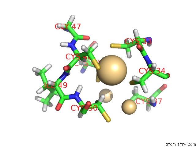
Mono view
Stereo pair view
|
A full contact list of Cadmium with other atoms in the Cd binding
site number 2 of The Three-Dimensional Structure of Human [113CD7] Metallothionein-2 in Solution Determined By Nuclear Magnetic Resonance Spectroscopy within 5.0Å range:
probe
|
atom
|
residue
|
distance (Å)
|
B
|
Occ
|
A:Cd105
b:1.0
occ:1.00
|
SG
|
A:CYS34
|
2.5
|
1.1
|
1.0
|
SG
|
A:CYS33
|
2.6
|
1.4
|
1.0
|
SG
|
A:CYS48
|
2.7
|
1.6
|
1.0
|
SG
|
A:CYS44
|
2.7
|
1.7
|
1.0
|
HB3
|
A:CYS44
|
2.9
|
1.7
|
1.0
|
HA
|
A:CYS48
|
3.0
|
0.6
|
1.0
|
HB3
|
A:CYS33
|
3.0
|
1.2
|
1.0
|
HB3
|
A:CYS50
|
3.0
|
1.2
|
1.0
|
HB3
|
A:CYS34
|
3.2
|
1.1
|
1.0
|
HB2
|
A:CYS48
|
3.3
|
1.4
|
1.0
|
CB
|
A:CYS33
|
3.4
|
1.3
|
1.0
|
CB
|
A:CYS44
|
3.4
|
1.1
|
1.0
|
CB
|
A:CYS48
|
3.4
|
1.0
|
1.0
|
CB
|
A:CYS34
|
3.4
|
1.1
|
1.0
|
HB2
|
A:CYS50
|
3.7
|
1.2
|
1.0
|
CA
|
A:CYS48
|
3.7
|
0.7
|
1.0
|
C
|
A:CYS33
|
3.8
|
0.9
|
1.0
|
H
|
A:CYS50
|
3.8
|
0.5
|
1.0
|
N
|
A:CYS34
|
3.9
|
0.9
|
1.0
|
CB
|
A:CYS50
|
3.9
|
0.8
|
1.0
|
O
|
A:CYS33
|
4.0
|
1.1
|
1.0
|
HB2
|
A:CYS44
|
4.0
|
2.2
|
1.0
|
H
|
A:CYS34
|
4.1
|
1.0
|
1.0
|
CA
|
A:CYS33
|
4.2
|
0.9
|
1.0
|
HB2
|
A:CYS37
|
4.2
|
1.5
|
1.0
|
CD
|
A:CD107
|
4.2
|
0.9
|
1.0
|
CA
|
A:CYS34
|
4.2
|
0.9
|
1.0
|
HB2
|
A:CYS33
|
4.3
|
1.6
|
1.0
|
HB2
|
A:CYS34
|
4.3
|
1.1
|
1.0
|
HA
|
A:CYS44
|
4.4
|
0.9
|
1.0
|
O
|
A:GLY47
|
4.4
|
1.6
|
1.0
|
HB3
|
A:CYS48
|
4.4
|
1.1
|
1.0
|
C
|
A:CYS48
|
4.5
|
0.7
|
1.0
|
CD
|
A:CD106
|
4.5
|
1.4
|
1.0
|
CA
|
A:CYS44
|
4.5
|
0.9
|
1.0
|
N
|
A:CYS50
|
4.6
|
0.6
|
1.0
|
H
|
A:ILE49
|
4.7
|
0.7
|
1.0
|
HA
|
A:CYS34
|
4.7
|
1.0
|
1.0
|
HA
|
A:CYS33
|
4.7
|
1.0
|
1.0
|
N
|
A:ILE49
|
4.8
|
0.6
|
1.0
|
CA
|
A:CYS50
|
4.8
|
0.6
|
1.0
|
N
|
A:CYS48
|
4.9
|
0.6
|
1.0
|
|
Cadmium binding site 3 out
of 4 in 1mhu
Go back to
Cadmium Binding Sites List in 1mhu
Cadmium binding site 3 out
of 4 in the The Three-Dimensional Structure of Human [113CD7] Metallothionein-2 in Solution Determined By Nuclear Magnetic Resonance Spectroscopy
Mono view
Stereo pair view
|
A full contact list of Cadmium with other atoms in the Cd binding
site number 3 of The Three-Dimensional Structure of Human [113CD7] Metallothionein-2 in Solution Determined By Nuclear Magnetic Resonance Spectroscopy within 5.0Å range:
probe
|
atom
|
residue
|
distance (Å)
|
B
|
Occ
|
A:Cd106
b:1.4
occ:1.00
|
SG
|
A:CYS44
|
2.4
|
1.7
|
1.0
|
SG
|
A:CYS37
|
2.6
|
1.7
|
1.0
|
SG
|
A:CYS41
|
2.7
|
2.8
|
1.0
|
SG
|
A:CYS60
|
2.7
|
1.4
|
1.0
|
HB2
|
A:CYS44
|
3.0
|
2.2
|
1.0
|
HB2
|
A:LYS43
|
3.1
|
3.4
|
1.0
|
HB3
|
A:CYS41
|
3.1
|
1.9
|
1.0
|
HB2
|
A:CYS50
|
3.1
|
1.2
|
1.0
|
HB3
|
A:CYS60
|
3.2
|
1.6
|
1.0
|
HB3
|
A:CYS37
|
3.3
|
1.7
|
1.0
|
CB
|
A:CYS44
|
3.3
|
1.1
|
1.0
|
CB
|
A:CYS60
|
3.4
|
1.4
|
1.0
|
CB
|
A:CYS37
|
3.4
|
1.0
|
1.0
|
CB
|
A:CYS41
|
3.4
|
0.9
|
1.0
|
HB2
|
A:CYS37
|
3.5
|
1.5
|
1.0
|
HB2
|
A:CYS60
|
3.6
|
1.2
|
1.0
|
O
|
A:LYS43
|
3.7
|
2.9
|
1.0
|
C
|
A:LYS43
|
3.7
|
1.7
|
1.0
|
HB2
|
A:CYS41
|
3.8
|
2.4
|
1.0
|
CB
|
A:LYS43
|
3.9
|
3.0
|
1.0
|
N
|
A:CYS44
|
4.0
|
0.9
|
1.0
|
HB3
|
A:LYS43
|
4.0
|
3.4
|
1.0
|
HB3
|
A:CYS44
|
4.1
|
1.7
|
1.0
|
CB
|
A:CYS50
|
4.2
|
0.8
|
1.0
|
CA
|
A:CYS44
|
4.2
|
0.9
|
1.0
|
CD
|
A:CD107
|
4.2
|
0.9
|
1.0
|
CA
|
A:LYS43
|
4.3
|
1.9
|
1.0
|
CD
|
A:CD101
|
4.3
|
1.0
|
1.0
|
H
|
A:LYS43
|
4.4
|
1.3
|
1.0
|
H
|
A:CYS44
|
4.4
|
1.5
|
1.0
|
CD
|
A:CD105
|
4.5
|
1.0
|
1.0
|
SG
|
A:CYS34
|
4.5
|
1.1
|
1.0
|
HB3
|
A:CYS50
|
4.5
|
1.2
|
1.0
|
HA
|
A:CYS44
|
4.6
|
0.9
|
1.0
|
N
|
A:LYS43
|
4.7
|
1.0
|
1.0
|
CA
|
A:CYS41
|
4.8
|
1.1
|
1.0
|
HD2
|
A:PRO38
|
4.8
|
1.2
|
1.0
|
CA
|
A:CYS60
|
4.8
|
1.6
|
1.0
|
CA
|
A:CYS37
|
4.8
|
0.9
|
1.0
|
SG
|
A:CYS50
|
4.8
|
0.6
|
1.0
|
O
|
A:CYS41
|
4.9
|
2.5
|
1.0
|
HG13
|
A:ILE49
|
5.0
|
3.5
|
1.0
|
|
Cadmium binding site 4 out
of 4 in 1mhu
Go back to
Cadmium Binding Sites List in 1mhu
Cadmium binding site 4 out
of 4 in the The Three-Dimensional Structure of Human [113CD7] Metallothionein-2 in Solution Determined By Nuclear Magnetic Resonance Spectroscopy
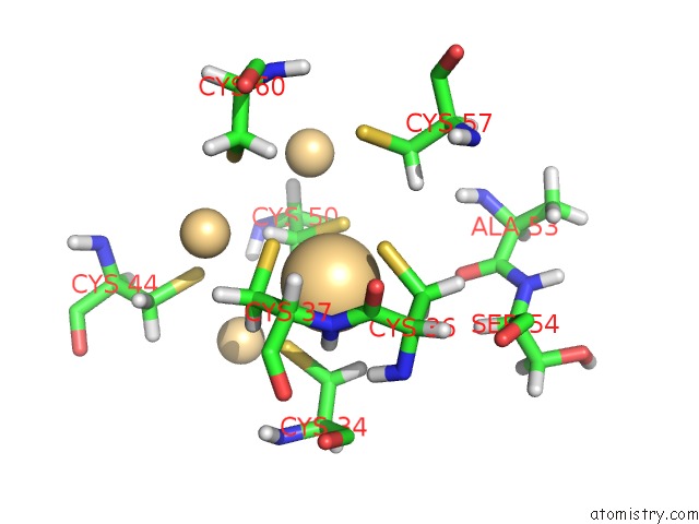
Mono view
Stereo pair view
|
A full contact list of Cadmium with other atoms in the Cd binding
site number 4 of The Three-Dimensional Structure of Human [113CD7] Metallothionein-2 in Solution Determined By Nuclear Magnetic Resonance Spectroscopy within 5.0Å range:
probe
|
atom
|
residue
|
distance (Å)
|
B
|
Occ
|
A:Cd107
b:0.9
occ:1.00
|
SG
|
A:CYS36
|
2.5
|
3.2
|
1.0
|
SG
|
A:CYS34
|
2.5
|
1.1
|
1.0
|
SG
|
A:CYS50
|
2.7
|
0.6
|
1.0
|
SG
|
A:CYS37
|
2.7
|
1.7
|
1.0
|
H
|
A:CYS37
|
2.8
|
1.0
|
1.0
|
HB2
|
A:CYS34
|
3.0
|
1.1
|
1.0
|
HB3
|
A:CYS36
|
3.0
|
1.4
|
1.0
|
HB3
|
A:CYS50
|
3.2
|
1.2
|
1.0
|
HB2
|
A:CYS37
|
3.2
|
1.5
|
1.0
|
N
|
A:CYS37
|
3.2
|
0.9
|
1.0
|
CB
|
A:CYS50
|
3.3
|
0.8
|
1.0
|
CB
|
A:CYS36
|
3.3
|
1.4
|
1.0
|
CB
|
A:CYS34
|
3.3
|
1.1
|
1.0
|
HB2
|
A:CYS50
|
3.3
|
1.2
|
1.0
|
CB
|
A:CYS37
|
3.4
|
1.0
|
1.0
|
H
|
A:CYS36
|
3.6
|
1.1
|
1.0
|
HB3
|
A:CYS34
|
3.8
|
1.1
|
1.0
|
C
|
A:CYS36
|
3.8
|
1.4
|
1.0
|
CA
|
A:CYS37
|
3.9
|
0.9
|
1.0
|
CA
|
A:CYS36
|
3.9
|
1.2
|
1.0
|
HB3
|
A:CYS57
|
4.1
|
0.8
|
1.0
|
N
|
A:CYS36
|
4.1
|
1.1
|
1.0
|
CD
|
A:CD105
|
4.2
|
1.0
|
1.0
|
CD
|
A:CD106
|
4.2
|
1.4
|
1.0
|
HB2
|
A:CYS36
|
4.2
|
1.5
|
1.0
|
CD
|
A:CD101
|
4.3
|
1.0
|
1.0
|
HA
|
A:CYS37
|
4.3
|
1.1
|
1.0
|
HB3
|
A:CYS37
|
4.4
|
1.7
|
1.0
|
CA
|
A:CYS34
|
4.6
|
0.9
|
1.0
|
SG
|
A:CYS44
|
4.7
|
1.7
|
1.0
|
O
|
A:ALA53
|
4.7
|
3.6
|
1.0
|
O
|
A:CYS36
|
4.7
|
2.2
|
1.0
|
CA
|
A:CYS50
|
4.8
|
0.6
|
1.0
|
HB2
|
A:CYS60
|
4.8
|
1.2
|
1.0
|
C
|
A:CYS34
|
4.9
|
1.0
|
1.0
|
SG
|
A:CYS57
|
4.9
|
1.2
|
1.0
|
CB
|
A:CYS57
|
4.9
|
0.9
|
1.0
|
HA
|
A:CYS36
|
5.0
|
1.2
|
1.0
|
HA
|
A:SER54
|
5.0
|
3.8
|
1.0
|
|
Reference:
B.A.Messerle,
A.Schaffer,
M.Vasak,
J.H.Kagi,
K.Wuthrich.
Three-Dimensional Structure of Human [113CD7]Metallothionein-2 in Solution Determined By Nuclear Magnetic Resonance Spectroscopy. J.Mol.Biol. V. 214 765 1990.
ISSN: ISSN 0022-2836
PubMed: 2388267
DOI: 10.1016/0022-2836(90)90291-S
Page generated: Fri Jul 19 13:52:15 2024
|