Cadmium »
PDB 1qvg-1vqo »
1rq1 »
Cadmium in PDB 1rq1: Structure of ERO1P, Source of Disulfide Bonds For Oxidative Protein Folding in the Cell
Protein crystallography data
The structure of Structure of ERO1P, Source of Disulfide Bonds For Oxidative Protein Folding in the Cell, PDB code: 1rq1
was solved by
E.Gross,
D.B.Kastner,
C.A.Kaiser,
D.Fass,
with X-Ray Crystallography technique. A brief refinement statistics is given in the table below:
| Resolution Low / High (Å) | 50.00 / 2.80 |
| Space group | P 62 |
| Cell size a, b, c (Å), α, β, γ (°) | 106.200, 106.200, 124.300, 90.00, 90.00, 120.00 |
| R / Rfree (%) | 20.6 / 23.8 |
Cadmium Binding Sites:
The binding sites of Cadmium atom in the Structure of ERO1P, Source of Disulfide Bonds For Oxidative Protein Folding in the Cell
(pdb code 1rq1). This binding sites where shown within
5.0 Angstroms radius around Cadmium atom.
In total 2 binding sites of Cadmium where determined in the Structure of ERO1P, Source of Disulfide Bonds For Oxidative Protein Folding in the Cell, PDB code: 1rq1:
Jump to Cadmium binding site number: 1; 2;
In total 2 binding sites of Cadmium where determined in the Structure of ERO1P, Source of Disulfide Bonds For Oxidative Protein Folding in the Cell, PDB code: 1rq1:
Jump to Cadmium binding site number: 1; 2;
Cadmium binding site 1 out of 2 in 1rq1
Go back to
Cadmium binding site 1 out
of 2 in the Structure of ERO1P, Source of Disulfide Bonds For Oxidative Protein Folding in the Cell
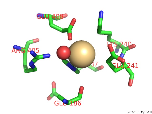
Mono view
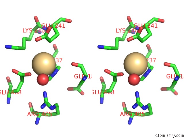
Stereo pair view
Mono view
Stereo pair view
A full contact list of Cadmium with other atoms in the Cd binding
site number 1 of Structure of ERO1P, Source of Disulfide Bonds For Oxidative Protein Folding in the Cell within 5.0Å range:
|
Cadmium binding site 2 out of 2 in 1rq1
Go back to
Cadmium binding site 2 out
of 2 in the Structure of ERO1P, Source of Disulfide Bonds For Oxidative Protein Folding in the Cell
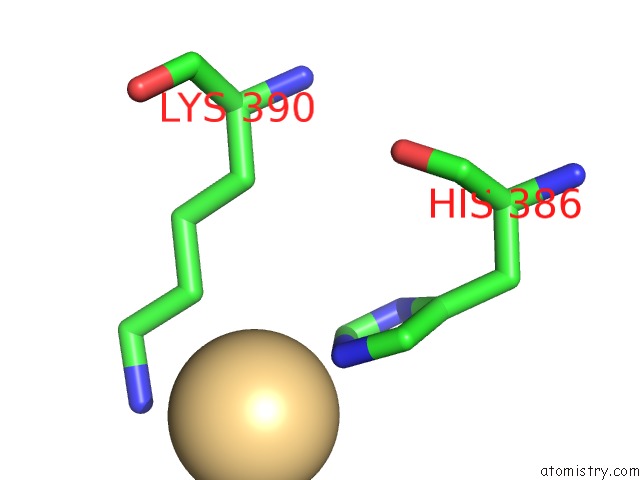
Mono view
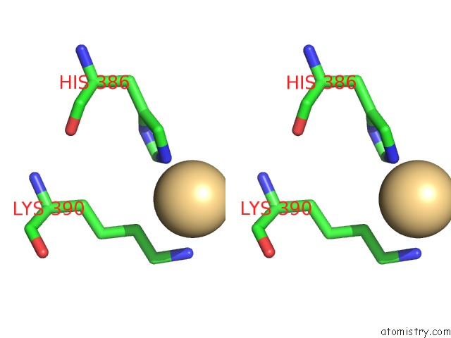
Stereo pair view
Mono view
Stereo pair view
A full contact list of Cadmium with other atoms in the Cd binding
site number 2 of Structure of ERO1P, Source of Disulfide Bonds For Oxidative Protein Folding in the Cell within 5.0Å range:
|
Reference:
E.Gross,
D.B.Kastner,
C.A.Kaiser,
D.Fass.
Structure of ERO1P, Source of Disulfide Bonds For Oxidative Protein Folding in the Cell. Cell(Cambridge,Mass.) V. 117 601 2004.
ISSN: ISSN 0092-8674
PubMed: 15163408
DOI: 10.1016/S0092-8674(04)00418-0
Page generated: Thu Jul 10 11:18:52 2025
ISSN: ISSN 0092-8674
PubMed: 15163408
DOI: 10.1016/S0092-8674(04)00418-0
Last articles
Cl in 1R08Cl in 1QZ9
Cl in 1QYE
Cl in 1QY8
Cl in 1QSG
Cl in 1QUO
Cl in 1QUL
Cl in 1QUH
Cl in 1QUJ
Cl in 1QTK