Cadmium »
PDB 1vqp-1yjw »
1x3f »
Cadmium in PDB 1x3f: Crystal Structure of the Single-Stranded Dna-Binding Protein From Mycobacterium Smegmatis
Protein crystallography data
The structure of Crystal Structure of the Single-Stranded Dna-Binding Protein From Mycobacterium Smegmatis, PDB code: 1x3f
was solved by
K.Saikrishnan,
G.P.Manjunath,
P.Singh,
J.Jeyakanthan,
Z.Dauter,
K.Sekar,
K.Muniyappa,
M.Vijayan,
with X-Ray Crystallography technique. A brief refinement statistics is given in the table below:
| Resolution Low / High (Å) | 20.00 / 2.70 |
| Space group | P 31 2 1 |
| Cell size a, b, c (Å), α, β, γ (°) | 78.014, 78.014, 71.018, 90.00, 90.00, 120.00 |
| R / Rfree (%) | 20 / 27.5 |
Cadmium Binding Sites:
The binding sites of Cadmium atom in the Crystal Structure of the Single-Stranded Dna-Binding Protein From Mycobacterium Smegmatis
(pdb code 1x3f). This binding sites where shown within
5.0 Angstroms radius around Cadmium atom.
In total 2 binding sites of Cadmium where determined in the Crystal Structure of the Single-Stranded Dna-Binding Protein From Mycobacterium Smegmatis, PDB code: 1x3f:
Jump to Cadmium binding site number: 1; 2;
In total 2 binding sites of Cadmium where determined in the Crystal Structure of the Single-Stranded Dna-Binding Protein From Mycobacterium Smegmatis, PDB code: 1x3f:
Jump to Cadmium binding site number: 1; 2;
Cadmium binding site 1 out of 2 in 1x3f
Go back to
Cadmium binding site 1 out
of 2 in the Crystal Structure of the Single-Stranded Dna-Binding Protein From Mycobacterium Smegmatis
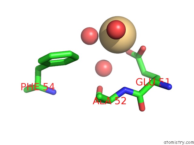
Mono view
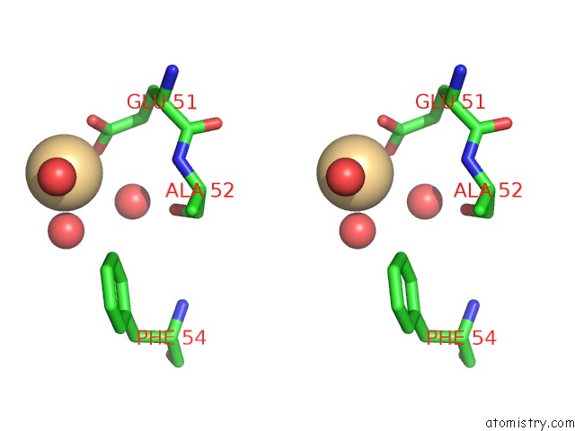
Stereo pair view
Mono view
Stereo pair view
A full contact list of Cadmium with other atoms in the Cd binding
site number 1 of Crystal Structure of the Single-Stranded Dna-Binding Protein From Mycobacterium Smegmatis within 5.0Å range:
|
Cadmium binding site 2 out of 2 in 1x3f
Go back to
Cadmium binding site 2 out
of 2 in the Crystal Structure of the Single-Stranded Dna-Binding Protein From Mycobacterium Smegmatis
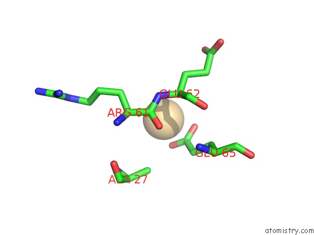
Mono view
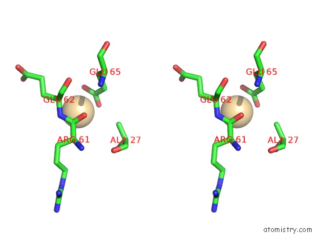
Stereo pair view
Mono view
Stereo pair view
A full contact list of Cadmium with other atoms in the Cd binding
site number 2 of Crystal Structure of the Single-Stranded Dna-Binding Protein From Mycobacterium Smegmatis within 5.0Å range:
|
Reference:
K.Saikrishnan,
G.P.Manjunath,
P.Singh,
J.Jeyakanthan,
Z.Dauter,
K.Sekar,
K.Muniyappa,
M.Vijayan.
Structure of Mycobacterium Smegmatis Single-Stranded Dna-Binding Protein and A Comparative Study Involving Homologus Ssbs: Biological Implications of Structural Plasticity and Variability in Quaternary Association. Acta Crystallogr.,Sect.D V. 61 1140 2005.
ISSN: ISSN 0907-4449
PubMed: 16041080
DOI: 10.1107/S0907444905016896
Page generated: Thu Jul 10 11:32:15 2025
ISSN: ISSN 0907-4449
PubMed: 16041080
DOI: 10.1107/S0907444905016896
Last articles
Fe in 2YXOFe in 2YRS
Fe in 2YXC
Fe in 2YNM
Fe in 2YVJ
Fe in 2YP1
Fe in 2YU2
Fe in 2YU1
Fe in 2YQB
Fe in 2YOO