Cadmium »
PDB 1vqp-1yjw »
1xmk »
Cadmium in PDB 1xmk: The Crystal Structure of the Zb Domain From the Rna Editing Enzyme ADAR1
Protein crystallography data
The structure of The Crystal Structure of the Zb Domain From the Rna Editing Enzyme ADAR1, PDB code: 1xmk
was solved by
A.Athanasiadis,
D.Placido,
S.Maas,
B.A.Brown Ii,
K.Lowenhaupt,
A.Rich,
with X-Ray Crystallography technique. A brief refinement statistics is given in the table below:
| Resolution Low / High (Å) | 10.00 / 0.97 |
| Space group | P 21 21 21 |
| Cell size a, b, c (Å), α, β, γ (°) | 35.557, 43.526, 45.471, 90.00, 90.00, 90.00 |
| R / Rfree (%) | 14.5 / 18.3 |
Other elements in 1xmk:
The structure of The Crystal Structure of the Zb Domain From the Rna Editing Enzyme ADAR1 also contains other interesting chemical elements:
| Nickel | (Ni) | 1 atom |
| Chlorine | (Cl) | 2 atoms |
Cadmium Binding Sites:
The binding sites of Cadmium atom in the The Crystal Structure of the Zb Domain From the Rna Editing Enzyme ADAR1
(pdb code 1xmk). This binding sites where shown within
5.0 Angstroms radius around Cadmium atom.
In total 2 binding sites of Cadmium where determined in the The Crystal Structure of the Zb Domain From the Rna Editing Enzyme ADAR1, PDB code: 1xmk:
Jump to Cadmium binding site number: 1; 2;
In total 2 binding sites of Cadmium where determined in the The Crystal Structure of the Zb Domain From the Rna Editing Enzyme ADAR1, PDB code: 1xmk:
Jump to Cadmium binding site number: 1; 2;
Cadmium binding site 1 out of 2 in 1xmk
Go back to
Cadmium binding site 1 out
of 2 in the The Crystal Structure of the Zb Domain From the Rna Editing Enzyme ADAR1
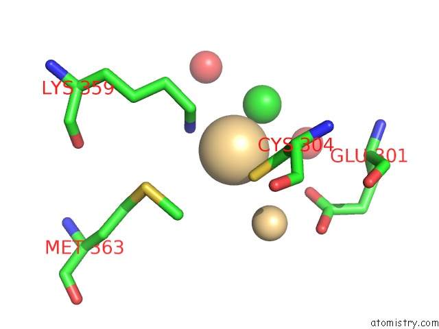
Mono view
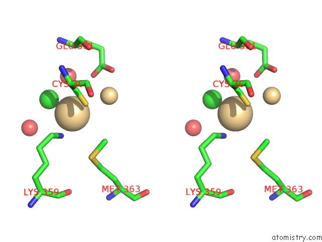
Stereo pair view
Mono view
Stereo pair view
A full contact list of Cadmium with other atoms in the Cd binding
site number 1 of The Crystal Structure of the Zb Domain From the Rna Editing Enzyme ADAR1 within 5.0Å range:
|
Cadmium binding site 2 out of 2 in 1xmk
Go back to
Cadmium binding site 2 out
of 2 in the The Crystal Structure of the Zb Domain From the Rna Editing Enzyme ADAR1
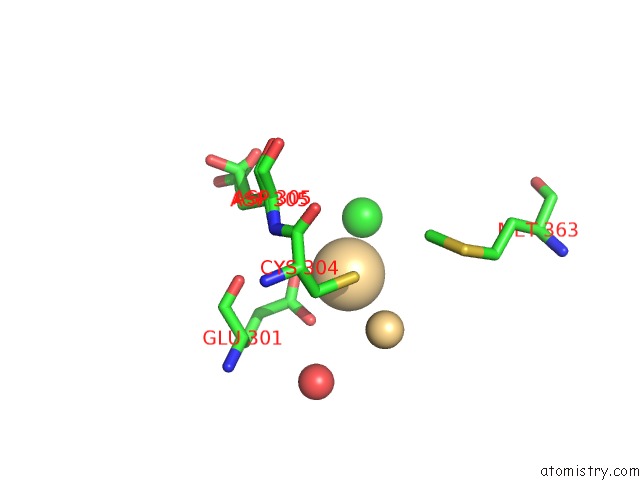
Mono view
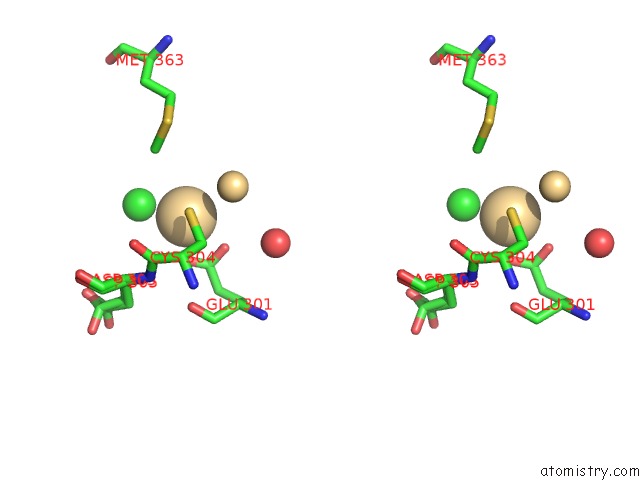
Stereo pair view
Mono view
Stereo pair view
A full contact list of Cadmium with other atoms in the Cd binding
site number 2 of The Crystal Structure of the Zb Domain From the Rna Editing Enzyme ADAR1 within 5.0Å range:
|
Reference:
A.Athanasiadis,
D.Placido,
S.Maas,
B.A.Brown Ii,
K.Lowenhaupt,
A.Rich.
The Crystal Structure of the Z[Beta] Domain of the Rna-Editing Enzyme ADAR1 Reveals Distinct Conserved Surfaces Among Z-Domains. J.Mol.Biol. V. 351 496 2005.
ISSN: ISSN 0022-2836
PubMed: 16023667
DOI: 10.1016/J.JMB.2005.06.028
Page generated: Thu Jul 10 11:32:38 2025
ISSN: ISSN 0022-2836
PubMed: 16023667
DOI: 10.1016/J.JMB.2005.06.028
Last articles
Fe in 2YXOFe in 2YRS
Fe in 2YXC
Fe in 2YNM
Fe in 2YVJ
Fe in 2YP1
Fe in 2YU2
Fe in 2YU1
Fe in 2YQB
Fe in 2YOO