Cadmium »
PDB 1ykq-2eik »
2c3g »
Cadmium in PDB 2c3g: Structure of CBM26 From Bacillus Halodurans Amylase
Protein crystallography data
The structure of Structure of CBM26 From Bacillus Halodurans Amylase, PDB code: 2c3g
was solved by
A.B.Boraston,
M.Healey,
J.Klassen,
E.Ficko-Blean,
A.Lammerts Van Bueren,
V.Law,
with X-Ray Crystallography technique. A brief refinement statistics is given in the table below:
| Resolution Low / High (Å) | 20.00 / 2.0 |
| Space group | P 42 21 2 |
| Cell size a, b, c (Å), α, β, γ (°) | 49.286, 49.286, 86.760, 90.00, 90.00, 90.00 |
| R / Rfree (%) | 22.6 / 30.2 |
Cadmium Binding Sites:
The binding sites of Cadmium atom in the Structure of CBM26 From Bacillus Halodurans Amylase
(pdb code 2c3g). This binding sites where shown within
5.0 Angstroms radius around Cadmium atom.
In total 4 binding sites of Cadmium where determined in the Structure of CBM26 From Bacillus Halodurans Amylase, PDB code: 2c3g:
Jump to Cadmium binding site number: 1; 2; 3; 4;
In total 4 binding sites of Cadmium where determined in the Structure of CBM26 From Bacillus Halodurans Amylase, PDB code: 2c3g:
Jump to Cadmium binding site number: 1; 2; 3; 4;
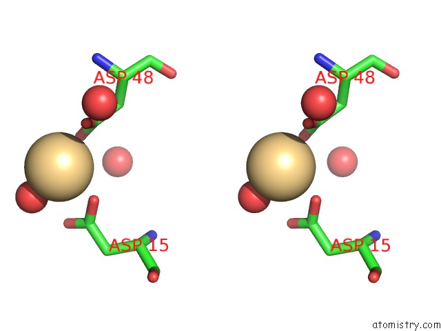
Cadmium binding site 1 out of 4 in 2c3g
Go back to
Cadmium binding site 1 out
of 4 in the Structure of CBM26 From Bacillus Halodurans Amylase

Mono view
Stereo pair view
Mono view
Stereo pair view
A full contact list of Cadmium with other atoms in the Cd binding
site number 1 of Structure of CBM26 From Bacillus Halodurans Amylase within 5.0Å range:
|
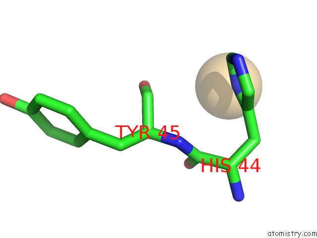
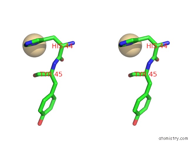
Cadmium binding site 2 out of 4 in 2c3g
Go back to
Cadmium binding site 2 out
of 4 in the Structure of CBM26 From Bacillus Halodurans Amylase

Mono view

Stereo pair view
Mono view
Stereo pair view
A full contact list of Cadmium with other atoms in the Cd binding
site number 2 of Structure of CBM26 From Bacillus Halodurans Amylase within 5.0Å range:
|
Cadmium binding site 3 out of 4 in 2c3g
Go back to
Cadmium binding site 3 out
of 4 in the Structure of CBM26 From Bacillus Halodurans Amylase

Mono view

Stereo pair view
Mono view
Stereo pair view
A full contact list of Cadmium with other atoms in the Cd binding
site number 3 of Structure of CBM26 From Bacillus Halodurans Amylase within 5.0Å range:
|
Cadmium binding site 4 out of 4 in 2c3g
Go back to
Cadmium binding site 4 out
of 4 in the Structure of CBM26 From Bacillus Halodurans Amylase
Mono view
Stereo pair view
Mono view
Stereo pair view
A full contact list of Cadmium with other atoms in the Cd binding
site number 4 of Structure of CBM26 From Bacillus Halodurans Amylase within 5.0Å range:
|
Reference:
A.B.Boraston,
M.Healey,
J.Klassen,
E.Ficko-Blean,
A.Lammerts Van Bueren,
V.Law.
A Structural and Functional Analysis of Alpha- Glucan Recognition By Family 25 and 26 Carbohydrate-Binding Modules Reveals A Conserved Mode of Starch Recognition J.Biol.Chem. V. 281 587 2006.
ISSN: ISSN 0021-9258
PubMed: 16230347
DOI: 10.1074/JBC.M509958200
Page generated: Thu Jul 10 11:43:12 2025
ISSN: ISSN 0021-9258
PubMed: 16230347
DOI: 10.1074/JBC.M509958200
Last articles
Fe in 2YXOFe in 2YRS
Fe in 2YXC
Fe in 2YNM
Fe in 2YVJ
Fe in 2YP1
Fe in 2YU2
Fe in 2YU1
Fe in 2YQB
Fe in 2YOO