Cadmium »
PDB 2wxt-3a05 »
2x7k »
Cadmium in PDB 2x7k: The Crystal Structure of PPIL1 in Complex with Cyclosporine A Suggests A Binding Mode For Skip
Enzymatic activity of The Crystal Structure of PPIL1 in Complex with Cyclosporine A Suggests A Binding Mode For Skip
All present enzymatic activity of The Crystal Structure of PPIL1 in Complex with Cyclosporine A Suggests A Binding Mode For Skip:
5.2.1.8;
5.2.1.8;
Protein crystallography data
The structure of The Crystal Structure of PPIL1 in Complex with Cyclosporine A Suggests A Binding Mode For Skip, PDB code: 2x7k
was solved by
C.M.Stegmann,
R.Luehrmann,
M.C.Wahl,
with X-Ray Crystallography technique. A brief refinement statistics is given in the table below:
| Resolution Low / High (Å) | 34.28 / 1.15 |
| Space group | P 21 21 2 |
| Cell size a, b, c (Å), α, β, γ (°) | 103.218, 35.701, 45.851, 90.00, 90.00, 90.00 |
| R / Rfree (%) | 12.8 / 14.9 |
Other elements in 2x7k:
The structure of The Crystal Structure of PPIL1 in Complex with Cyclosporine A Suggests A Binding Mode For Skip also contains other interesting chemical elements:
| Sodium | (Na) | 1 atom |
Cadmium Binding Sites:
The binding sites of Cadmium atom in the The Crystal Structure of PPIL1 in Complex with Cyclosporine A Suggests A Binding Mode For Skip
(pdb code 2x7k). This binding sites where shown within
5.0 Angstroms radius around Cadmium atom.
In total 2 binding sites of Cadmium where determined in the The Crystal Structure of PPIL1 in Complex with Cyclosporine A Suggests A Binding Mode For Skip, PDB code: 2x7k:
Jump to Cadmium binding site number: 1; 2;
In total 2 binding sites of Cadmium where determined in the The Crystal Structure of PPIL1 in Complex with Cyclosporine A Suggests A Binding Mode For Skip, PDB code: 2x7k:
Jump to Cadmium binding site number: 1; 2;
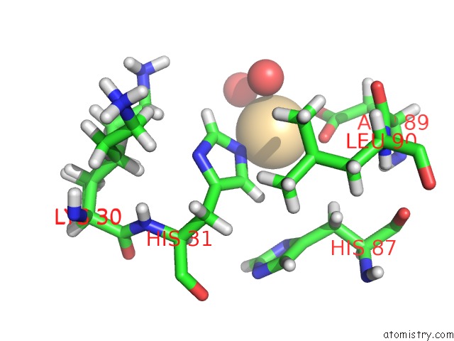
Cadmium binding site 1 out of 2 in 2x7k
Go back to
Cadmium binding site 1 out
of 2 in the The Crystal Structure of PPIL1 in Complex with Cyclosporine A Suggests A Binding Mode For Skip
Mono view

Stereo pair view
Mono view
Stereo pair view
A full contact list of Cadmium with other atoms in the Cd binding
site number 1 of The Crystal Structure of PPIL1 in Complex with Cyclosporine A Suggests A Binding Mode For Skip within 5.0Å range:
|
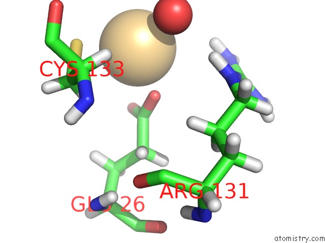
Cadmium binding site 2 out of 2 in 2x7k
Go back to
Cadmium binding site 2 out
of 2 in the The Crystal Structure of PPIL1 in Complex with Cyclosporine A Suggests A Binding Mode For Skip
Mono view

Stereo pair view
Mono view
Stereo pair view
A full contact list of Cadmium with other atoms in the Cd binding
site number 2 of The Crystal Structure of PPIL1 in Complex with Cyclosporine A Suggests A Binding Mode For Skip within 5.0Å range:
|
Reference:
C.M.Stegmann,
R.Luehrmann,
M.C.Wahl.
The Crystal Structure of PPIL1 Bound to Cyclosporine A Suggests A Binding Mode For A Linear Epitope of the Skip Protein. Plos One V. 5 13 2010.
ISSN: ESSN 1932-6203
PubMed: 20368803
DOI: 10.1371/JOURNAL.PONE.0010013
Page generated: Thu Jul 10 12:13:05 2025
ISSN: ESSN 1932-6203
PubMed: 20368803
DOI: 10.1371/JOURNAL.PONE.0010013
Last articles
Fe in 2YXOFe in 2YRS
Fe in 2YXC
Fe in 2YNM
Fe in 2YVJ
Fe in 2YP1
Fe in 2YU2
Fe in 2YU1
Fe in 2YQB
Fe in 2YOO