Cadmium »
PDB 3af7-3cme »
3c7m »
Cadmium in PDB 3c7m: Crystal Structure of Reduced Dsbl
Protein crystallography data
The structure of Crystal Structure of Reduced Dsbl, PDB code: 3c7m
was solved by
C.U.Stirnimann,
J.P.A.Grimshaw,
R.Glockshuber,
M.G.Grutter,
G.Capitani,
with X-Ray Crystallography technique. A brief refinement statistics is given in the table below:
| Resolution Low / High (Å) | 19.74 / 1.55 |
| Space group | P 1 21 1 |
| Cell size a, b, c (Å), α, β, γ (°) | 50.945, 66.634, 77.891, 90.00, 107.02, 90.00 |
| R / Rfree (%) | 16.4 / 18.9 |
Other elements in 3c7m:
The structure of Crystal Structure of Reduced Dsbl also contains other interesting chemical elements:
| Chlorine | (Cl) | 11 atoms |
Cadmium Binding Sites:
The binding sites of Cadmium atom in the Crystal Structure of Reduced Dsbl
(pdb code 3c7m). This binding sites where shown within
5.0 Angstroms radius around Cadmium atom.
In total 6 binding sites of Cadmium where determined in the Crystal Structure of Reduced Dsbl, PDB code: 3c7m:
Jump to Cadmium binding site number: 1; 2; 3; 4; 5; 6;
In total 6 binding sites of Cadmium where determined in the Crystal Structure of Reduced Dsbl, PDB code: 3c7m:
Jump to Cadmium binding site number: 1; 2; 3; 4; 5; 6;
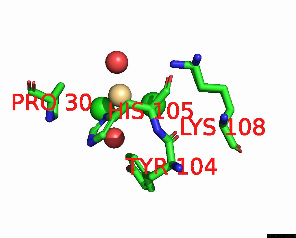
Cadmium binding site 1 out of 6 in 3c7m
Go back to
Cadmium binding site 1 out
of 6 in the Crystal Structure of Reduced Dsbl

Mono view

Stereo pair view
Mono view
Stereo pair view
A full contact list of Cadmium with other atoms in the Cd binding
site number 1 of Crystal Structure of Reduced Dsbl within 5.0Å range:
|
Cadmium binding site 2 out of 6 in 3c7m
Go back to
Cadmium binding site 2 out
of 6 in the Crystal Structure of Reduced Dsbl

Mono view

Stereo pair view
Mono view
Stereo pair view
A full contact list of Cadmium with other atoms in the Cd binding
site number 2 of Crystal Structure of Reduced Dsbl within 5.0Å range:
|
Cadmium binding site 3 out of 6 in 3c7m
Go back to
Cadmium binding site 3 out
of 6 in the Crystal Structure of Reduced Dsbl

Mono view

Stereo pair view
Mono view
Stereo pair view
A full contact list of Cadmium with other atoms in the Cd binding
site number 3 of Crystal Structure of Reduced Dsbl within 5.0Å range:
|
Cadmium binding site 4 out of 6 in 3c7m
Go back to
Cadmium binding site 4 out
of 6 in the Crystal Structure of Reduced Dsbl
Mono view

Stereo pair view
Mono view
Stereo pair view
A full contact list of Cadmium with other atoms in the Cd binding
site number 4 of Crystal Structure of Reduced Dsbl within 5.0Å range:
|
Cadmium binding site 5 out of 6 in 3c7m
Go back to
Cadmium binding site 5 out
of 6 in the Crystal Structure of Reduced Dsbl

Mono view

Stereo pair view
Mono view
Stereo pair view
A full contact list of Cadmium with other atoms in the Cd binding
site number 5 of Crystal Structure of Reduced Dsbl within 5.0Å range:
|
Cadmium binding site 6 out of 6 in 3c7m
Go back to
Cadmium binding site 6 out
of 6 in the Crystal Structure of Reduced Dsbl

Mono view

Stereo pair view
Mono view
Stereo pair view
A full contact list of Cadmium with other atoms in the Cd binding
site number 6 of Crystal Structure of Reduced Dsbl within 5.0Å range:
|
Reference:
J.P.Grimshaw,
C.U.Stirnimann,
M.S.Brozzo,
G.Malojcic,
M.G.Grutter,
G.Capitani,
R.Glockshuber.
Dsbl and Dsbi Form A Specific Dithiol Oxidase System For Periplasmic Arylsulfate Sulfotransferase in Uropathogenic Escherichia Coli. J.Mol.Biol. V. 380 667 2008.
ISSN: ISSN 0022-2836
PubMed: 18565543
DOI: 10.1016/J.JMB.2008.05.031
Page generated: Thu Jul 10 12:22:08 2025
ISSN: ISSN 0022-2836
PubMed: 18565543
DOI: 10.1016/J.JMB.2008.05.031
Last articles
Fe in 2YXOFe in 2YRS
Fe in 2YXC
Fe in 2YNM
Fe in 2YVJ
Fe in 2YP1
Fe in 2YU2
Fe in 2YU1
Fe in 2YQB
Fe in 2YOO