Cadmium »
PDB 3heq-3o7s »
3hrt »
Cadmium in PDB 3hrt: Crystal Structure of Scar with Bound CD2+
Protein crystallography data
The structure of Crystal Structure of Scar with Bound CD2+, PDB code: 3hrt
was solved by
K.E.Stoll,
W.E.Draper,
J.I.Kliegman,
M.V.Golynskiy,
R.A.T.Brew-Appiah,
H.K.Brown,
W.A.Breyer,
N.S.Jakubovics,
H.F.Jenkinson,
R.B.Brennan,
S.M.Cohen,
A.Glasfeld,
with X-Ray Crystallography technique. A brief refinement statistics is given in the table below:
| Resolution Low / High (Å) | 30.25 / 2.80 |
| Space group | P 41 21 2 |
| Cell size a, b, c (Å), α, β, γ (°) | 70.860, 70.860, 304.100, 90.00, 90.00, 90.00 |
| R / Rfree (%) | 23 / 28.2 |
Cadmium Binding Sites:
The binding sites of Cadmium atom in the Crystal Structure of Scar with Bound CD2+
(pdb code 3hrt). This binding sites where shown within
5.0 Angstroms radius around Cadmium atom.
In total 2 binding sites of Cadmium where determined in the Crystal Structure of Scar with Bound CD2+, PDB code: 3hrt:
Jump to Cadmium binding site number: 1; 2;
In total 2 binding sites of Cadmium where determined in the Crystal Structure of Scar with Bound CD2+, PDB code: 3hrt:
Jump to Cadmium binding site number: 1; 2;
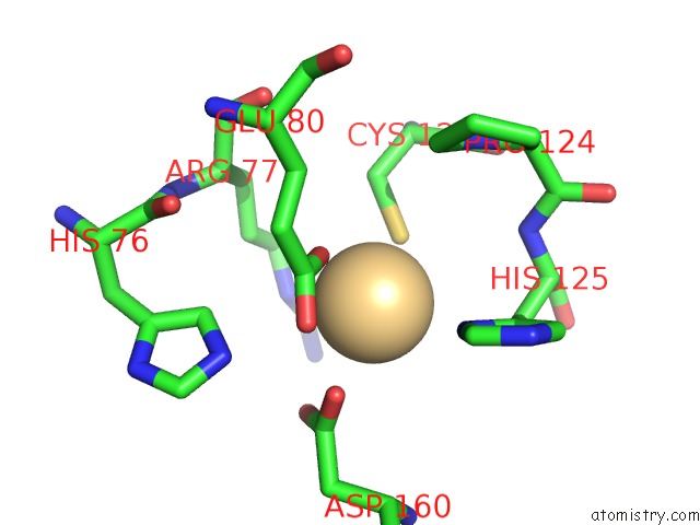
Cadmium binding site 1 out of 2 in 3hrt
Go back to
Cadmium binding site 1 out
of 2 in the Crystal Structure of Scar with Bound CD2+
Mono view
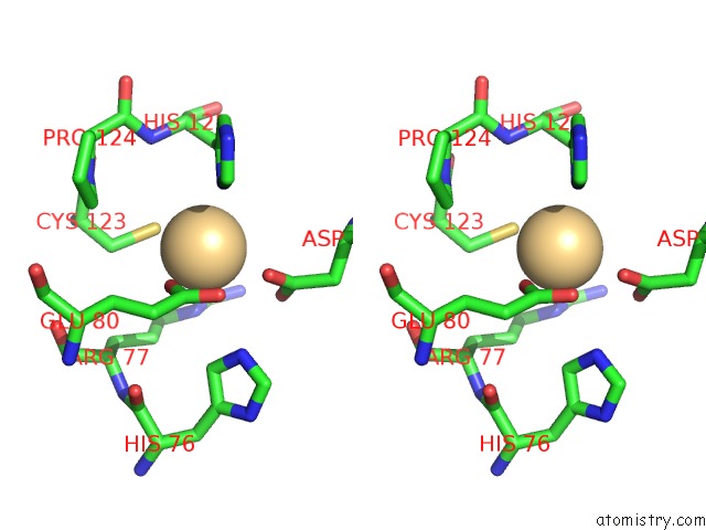
Stereo pair view
Mono view
Stereo pair view
A full contact list of Cadmium with other atoms in the Cd binding
site number 1 of Crystal Structure of Scar with Bound CD2+ within 5.0Å range:
|
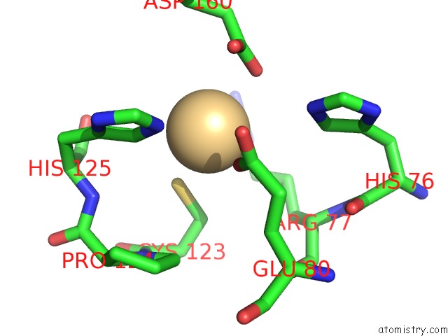
Cadmium binding site 2 out of 2 in 3hrt
Go back to
Cadmium binding site 2 out
of 2 in the Crystal Structure of Scar with Bound CD2+
Mono view
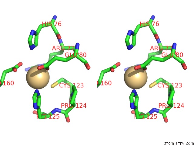
Stereo pair view
Mono view
Stereo pair view
A full contact list of Cadmium with other atoms in the Cd binding
site number 2 of Crystal Structure of Scar with Bound CD2+ within 5.0Å range:
|
Reference:
K.E.Stoll,
W.E.Draper,
J.I.Kliegman,
M.V.Golynskiy,
R.A.Brew-Appiah,
R.K.Phillips,
H.K.Brown,
W.A.Breyer,
N.S.Jakubovics,
H.F.Jenkinson,
R.G.Brennan,
S.M.Cohen,
A.Glasfeld.
Characterization and Structure of the Manganese-Responsive Transcriptional Regulator Scar. Biochemistry V. 48 10308 2009.
ISSN: ISSN 0006-2960
PubMed: 19795834
DOI: 10.1021/BI900980G
Page generated: Thu Jul 10 12:41:35 2025
ISSN: ISSN 0006-2960
PubMed: 19795834
DOI: 10.1021/BI900980G
Last articles
Cl in 7Z2GCl in 7Z2H
Cl in 7Z1W
Cl in 7YY9
Cl in 7Z1G
Cl in 7YZZ
Cl in 7Z1F
Cl in 7Z0V
Cl in 7YYG
Cl in 7YWK