Cadmium »
PDB 5wgr-6fpc »
5wro »
Cadmium in PDB 5wro: Crystal Structure of Drosophila Enolase
Enzymatic activity of Crystal Structure of Drosophila Enolase
All present enzymatic activity of Crystal Structure of Drosophila Enolase:
4.2.1.11;
4.2.1.11;
Protein crystallography data
The structure of Crystal Structure of Drosophila Enolase, PDB code: 5wro
was solved by
Z.Zhang,
Z.Shi,
with X-Ray Crystallography technique. A brief refinement statistics is given in the table below:
| Resolution Low / High (Å) | 38.33 / 2.02 |
| Space group | H 3 2 |
| Cell size a, b, c (Å), α, β, γ (°) | 118.820, 118.820, 229.792, 90.00, 90.00, 120.00 |
| R / Rfree (%) | 15.9 / 20 |
Other elements in 5wro:
The structure of Crystal Structure of Drosophila Enolase also contains other interesting chemical elements:
| Cobalt | (Co) | 1 atom |
| Chlorine | (Cl) | 1 atom |
Cadmium Binding Sites:
The binding sites of Cadmium atom in the Crystal Structure of Drosophila Enolase
(pdb code 5wro). This binding sites where shown within
5.0 Angstroms radius around Cadmium atom.
In total 2 binding sites of Cadmium where determined in the Crystal Structure of Drosophila Enolase, PDB code: 5wro:
Jump to Cadmium binding site number: 1; 2;
In total 2 binding sites of Cadmium where determined in the Crystal Structure of Drosophila Enolase, PDB code: 5wro:
Jump to Cadmium binding site number: 1; 2;
Cadmium binding site 1 out of 2 in 5wro
Go back to
Cadmium binding site 1 out
of 2 in the Crystal Structure of Drosophila Enolase
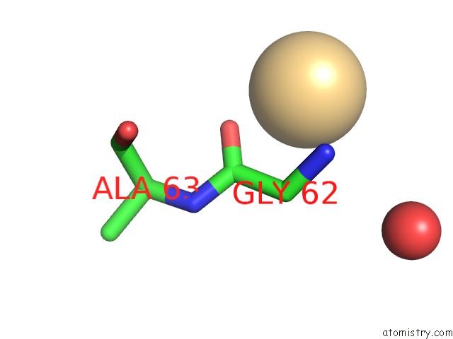
Mono view
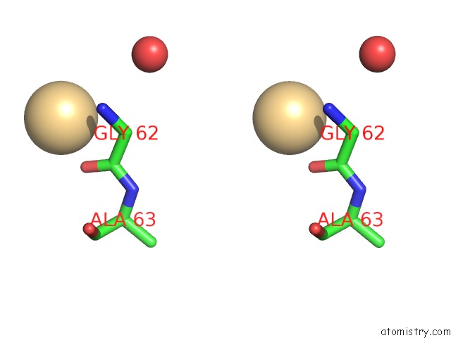
Stereo pair view
Mono view
Stereo pair view
A full contact list of Cadmium with other atoms in the Cd binding
site number 1 of Crystal Structure of Drosophila Enolase within 5.0Å range:
|
Cadmium binding site 2 out of 2 in 5wro
Go back to
Cadmium binding site 2 out
of 2 in the Crystal Structure of Drosophila Enolase
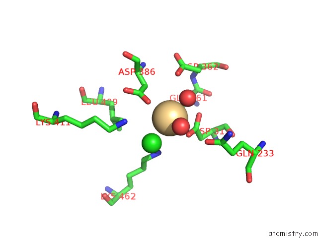
Mono view
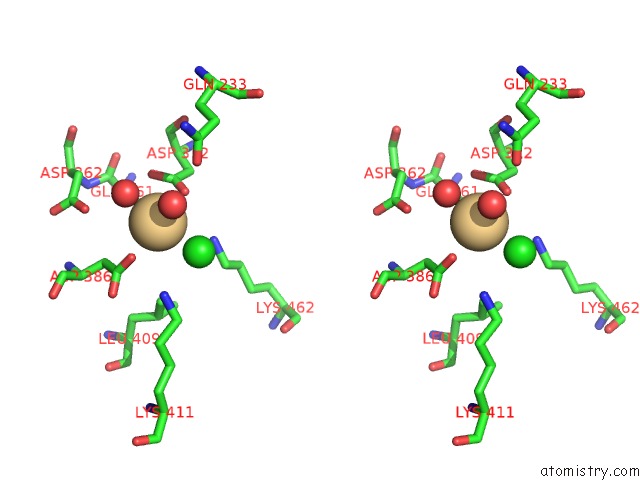
Stereo pair view
Mono view
Stereo pair view
A full contact list of Cadmium with other atoms in the Cd binding
site number 2 of Crystal Structure of Drosophila Enolase within 5.0Å range:
|
Reference:
C.Sun,
B.Xu,
X.Liu,
Z.Zhang,
Z.Su.
Crystal Structure of Enolase From Drosophila Melanogaster. Acta Crystallogr F Struct V. 73 228 2017BIOL Commun.
ISSN: ESSN 2053-230X
PubMed: 28368282
DOI: 10.1107/S2053230X17004022
Page generated: Thu Jul 10 14:44:49 2025
ISSN: ESSN 2053-230X
PubMed: 28368282
DOI: 10.1107/S2053230X17004022
Last articles
F in 7O2EF in 7O2B
F in 7NZU
F in 7O29
F in 7NZS
F in 7NZT
F in 7NY9
F in 7NY2
F in 7NYA
F in 7NZR