Cadmium »
PDB 1dpe-1gwg »
1gm6 »
Cadmium in PDB 1gm6: 3-D Structure of A Salivary Lipocalin From Boar
Protein crystallography data
The structure of 3-D Structure of A Salivary Lipocalin From Boar, PDB code: 1gm6
was solved by
S.Spinelli,
F.Vincent,
P.Pelosi,
M.Tegoni,
C.Cambillau,
with X-Ray Crystallography technique. A brief refinement statistics is given in the table below:
| Resolution Low / High (Å) | 14.53 / 2.13 |
| Space group | P 41 21 2 |
| Cell size a, b, c (Å), α, β, γ (°) | 70.112, 70.112, 71.750, 90.00, 90.00, 90.00 |
| R / Rfree (%) | 25.4 / 28.2 |
Cadmium Binding Sites:
The binding sites of Cadmium atom in the 3-D Structure of A Salivary Lipocalin From Boar
(pdb code 1gm6). This binding sites where shown within
5.0 Angstroms radius around Cadmium atom.
In total only one binding site of Cadmium was determined in the 3-D Structure of A Salivary Lipocalin From Boar, PDB code: 1gm6:
In total only one binding site of Cadmium was determined in the 3-D Structure of A Salivary Lipocalin From Boar, PDB code: 1gm6:
Cadmium binding site 1 out of 1 in 1gm6
Go back to
Cadmium binding site 1 out
of 1 in the 3-D Structure of A Salivary Lipocalin From Boar
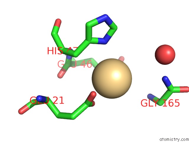
Mono view
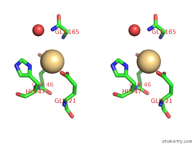
Stereo pair view
Mono view
Stereo pair view
A full contact list of Cadmium with other atoms in the Cd binding
site number 1 of 3-D Structure of A Salivary Lipocalin From Boar within 5.0Å range:
|
Reference:
S.Spinelli,
F.Vincent,
P.Pelosi,
M.Tegoni,
C.Cambillau.
Boar Salivary Lipocalin. Three-Dimensional X-Ray Structure and Androsterol/Androstenone Docking Simulations. Eur.J.Biochem. V. 269 2449 2002.
ISSN: ISSN 0014-2956
PubMed: 12027882
DOI: 10.1046/J.1432-1033.2002.02901.X
Page generated: Thu Jul 10 10:45:37 2025
ISSN: ISSN 0014-2956
PubMed: 12027882
DOI: 10.1046/J.1432-1033.2002.02901.X
Last articles
Na in 8VNHNa in 8VNG
Na in 8VNF
Na in 8VNE
Na in 8VNA
Na in 8VN9
Na in 8VN8
Na in 8VN7
Na in 8VN6
Na in 8VN4