Cadmium »
PDB 3af7-3cme »
3afp »
Cadmium in PDB 3afp: Crystal Structure of the Single-Stranded Dna Binding Protein From Mycobacterium Leprae (Form I)
Protein crystallography data
The structure of Crystal Structure of the Single-Stranded Dna Binding Protein From Mycobacterium Leprae (Form I), PDB code: 3afp
was solved by
P.S.Kaushal,
P.Singh,
A.Sharma,
K.Muniyappa,
M.Vijayan,
with X-Ray Crystallography technique. A brief refinement statistics is given in the table below:
| Resolution Low / High (Å) | 28.14 / 2.05 |
| Space group | P 31 2 1 |
| Cell size a, b, c (Å), α, β, γ (°) | 78.957, 78.957, 80.213, 90.00, 90.00, 120.00 |
| R / Rfree (%) | 20.4 / 23.2 |
Cadmium Binding Sites:
The binding sites of Cadmium atom in the Crystal Structure of the Single-Stranded Dna Binding Protein From Mycobacterium Leprae (Form I)
(pdb code 3afp). This binding sites where shown within
5.0 Angstroms radius around Cadmium atom.
In total only one binding site of Cadmium was determined in the Crystal Structure of the Single-Stranded Dna Binding Protein From Mycobacterium Leprae (Form I), PDB code: 3afp:
In total only one binding site of Cadmium was determined in the Crystal Structure of the Single-Stranded Dna Binding Protein From Mycobacterium Leprae (Form I), PDB code: 3afp:
Cadmium binding site 1 out of 1 in 3afp
Go back to
Cadmium binding site 1 out
of 1 in the Crystal Structure of the Single-Stranded Dna Binding Protein From Mycobacterium Leprae (Form I)
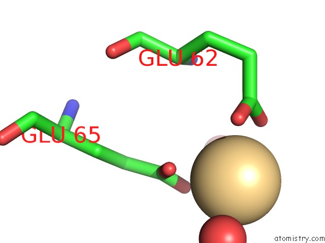
Mono view
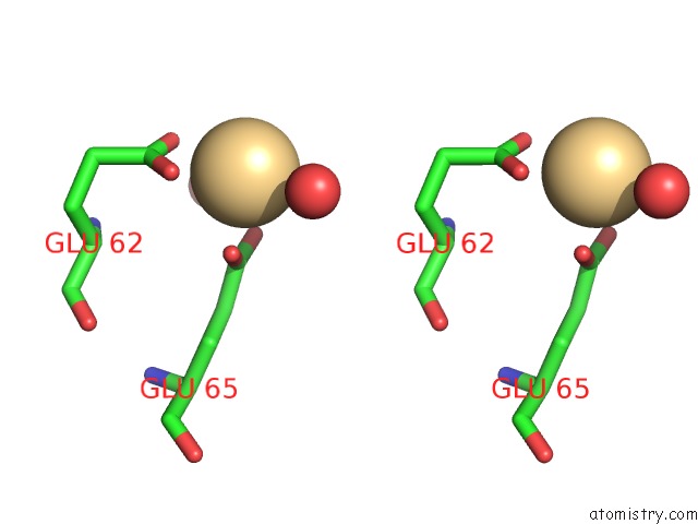
Stereo pair view
Mono view
Stereo pair view
A full contact list of Cadmium with other atoms in the Cd binding
site number 1 of Crystal Structure of the Single-Stranded Dna Binding Protein From Mycobacterium Leprae (Form I) within 5.0Å range:
|
Reference:
P.S.Kaushal,
P.Singh,
A.Sharma,
K.Muniyappa,
M.Vijayan.
X-Ray and Molecular-Dynamics Studies on Mycobacterium Leprae Single-Stranded Dna-Binding Protein and Comparison with Other Eubacterial Ssb Structures Acta Crystallogr.,Sect.D V. 66 1048 2010.
ISSN: ISSN 0907-4449
PubMed: 20944238
DOI: 10.1107/S0907444910032208
Page generated: Thu Jul 10 12:19:34 2025
ISSN: ISSN 0907-4449
PubMed: 20944238
DOI: 10.1107/S0907444910032208
Last articles
Na in 5MEWNa in 5MEJ
Na in 5MEH
Na in 5MDQ
Na in 5MDU
Na in 5MC3
Na in 5MB4
Na in 5MC1
Na in 5MC2
Na in 5M99