Cadmium »
PDB 7viq-8hyf »
8ghj »
Cadmium in PDB 8ghj: Crystal Structure of Human AQP2 T125M Mutant
Protein crystallography data
The structure of Crystal Structure of Human AQP2 T125M Mutant, PDB code: 8ghj
was solved by
S.Horsefield,
C.J.Hagstroemer,
with X-Ray Crystallography technique. A brief refinement statistics is given in the table below:
| Resolution Low / High (Å) | 49.50 / 3.90 |
| Space group | P 42 |
| Cell size a, b, c (Å), α, β, γ (°) | 118.31, 118.31, 90.4, 90, 90, 90 |
| R / Rfree (%) | 28.8 / 31.3 |
Cadmium Binding Sites:
The binding sites of Cadmium atom in the Crystal Structure of Human AQP2 T125M Mutant
(pdb code 8ghj). This binding sites where shown within
5.0 Angstroms radius around Cadmium atom.
In total 2 binding sites of Cadmium where determined in the Crystal Structure of Human AQP2 T125M Mutant, PDB code: 8ghj:
Jump to Cadmium binding site number: 1; 2;
In total 2 binding sites of Cadmium where determined in the Crystal Structure of Human AQP2 T125M Mutant, PDB code: 8ghj:
Jump to Cadmium binding site number: 1; 2;
Cadmium binding site 1 out of 2 in 8ghj
Go back to
Cadmium binding site 1 out
of 2 in the Crystal Structure of Human AQP2 T125M Mutant

Mono view
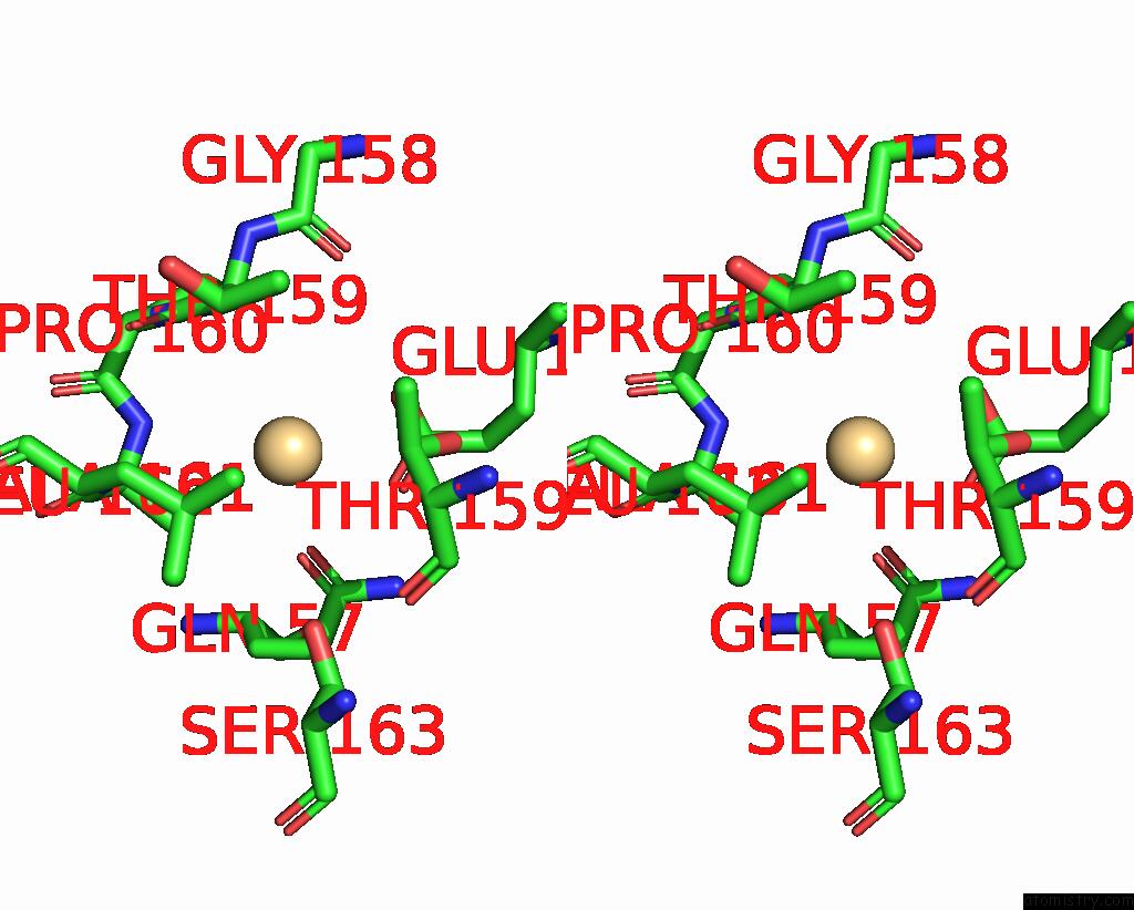
Stereo pair view
Mono view
Stereo pair view
A full contact list of Cadmium with other atoms in the Cd binding
site number 1 of Crystal Structure of Human AQP2 T125M Mutant within 5.0Å range:
|
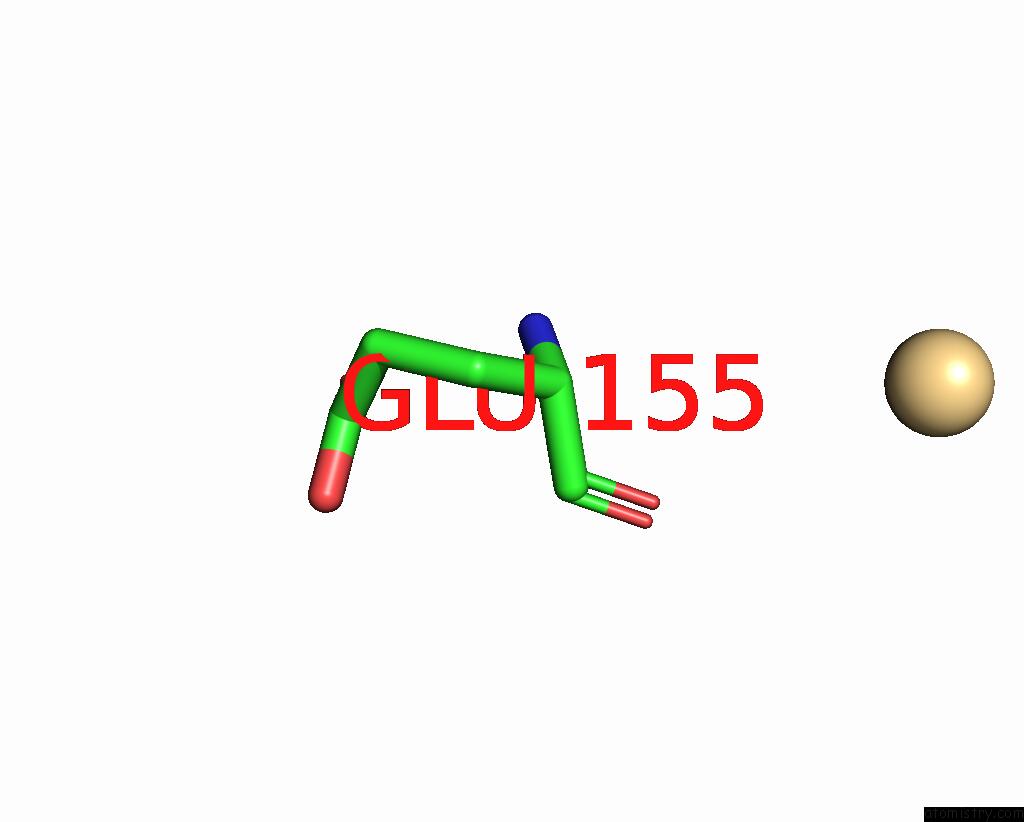
Cadmium binding site 2 out of 2 in 8ghj
Go back to
Cadmium binding site 2 out
of 2 in the Crystal Structure of Human AQP2 T125M Mutant
Mono view
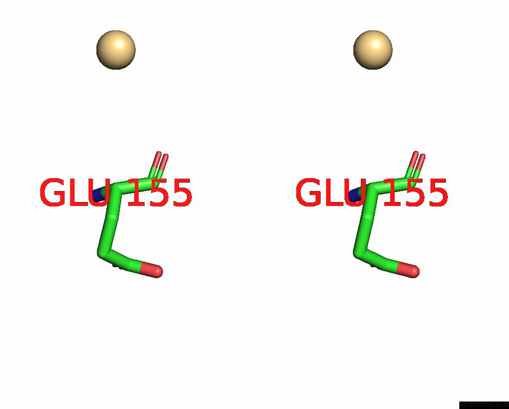
Stereo pair view
Mono view
Stereo pair view
A full contact list of Cadmium with other atoms in the Cd binding
site number 2 of Crystal Structure of Human AQP2 T125M Mutant within 5.0Å range:
|
Reference:
C.J.Hagstromer,
J.Hyld Steffen,
S.Kreida,
T.Al-Jubair,
A.Frick,
P.Gourdon,
S.Tornroth-Horsefield.
Structural and Functional Analysis of Aquaporin-2 Mutants Involved in Nephrogenic Diabetes Insipidus. Sci Rep V. 13 14674 2023.
ISSN: ESSN 2045-2322
PubMed: 37674034
DOI: 10.1038/S41598-023-41616-1
Page generated: Thu Jul 10 15:44:33 2025
ISSN: ESSN 2045-2322
PubMed: 37674034
DOI: 10.1038/S41598-023-41616-1
Last articles
Mg in 7DR0Mg in 7DR1
Mg in 7DU2
Mg in 7DSP
Mg in 7DSJ
Mg in 7DSI
Mg in 7DRP
Mg in 7DSH
Mg in 7DSA
Mg in 7DRX