Cadmium »
PDB 1mws-1qvf »
1oo2 »
Cadmium in PDB 1oo2: Crystal Structure of Transthyretin From Sparus Aurata
Protein crystallography data
The structure of Crystal Structure of Transthyretin From Sparus Aurata, PDB code: 1oo2
was solved by
N.Pasquato,
I.Ramazzina,
C.Folli,
R.Battistutta,
R.Berni,
G.Zanotti,
with X-Ray Crystallography technique. A brief refinement statistics is given in the table below:
| Resolution Low / High (Å) | 10.00 / 1.56 |
| Space group | C 1 2 1 |
| Cell size a, b, c (Å), α, β, γ (°) | 96.452, 65.622, 70.834, 90.00, 97.40, 90.00 |
| R / Rfree (%) | 19.7 / 22.4 |
Cadmium Binding Sites:
The binding sites of Cadmium atom in the Crystal Structure of Transthyretin From Sparus Aurata
(pdb code 1oo2). This binding sites where shown within
5.0 Angstroms radius around Cadmium atom.
In total 2 binding sites of Cadmium where determined in the Crystal Structure of Transthyretin From Sparus Aurata, PDB code: 1oo2:
Jump to Cadmium binding site number: 1; 2;
In total 2 binding sites of Cadmium where determined in the Crystal Structure of Transthyretin From Sparus Aurata, PDB code: 1oo2:
Jump to Cadmium binding site number: 1; 2;
Cadmium binding site 1 out of 2 in 1oo2
Go back to
Cadmium binding site 1 out
of 2 in the Crystal Structure of Transthyretin From Sparus Aurata
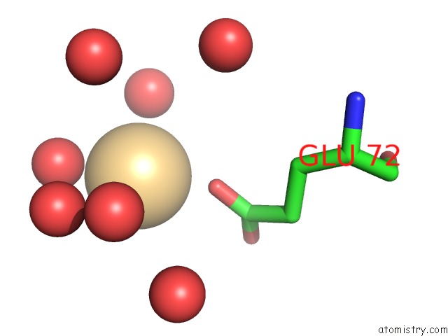
Mono view
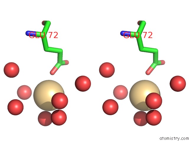
Stereo pair view
Mono view
Stereo pair view
A full contact list of Cadmium with other atoms in the Cd binding
site number 1 of Crystal Structure of Transthyretin From Sparus Aurata within 5.0Å range:
|
Cadmium binding site 2 out of 2 in 1oo2
Go back to
Cadmium binding site 2 out
of 2 in the Crystal Structure of Transthyretin From Sparus Aurata
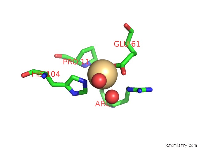
Mono view
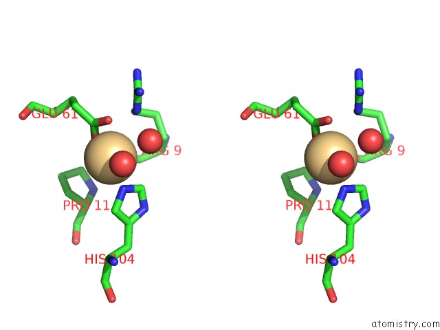
Stereo pair view
Mono view
Stereo pair view
A full contact list of Cadmium with other atoms in the Cd binding
site number 2 of Crystal Structure of Transthyretin From Sparus Aurata within 5.0Å range:
|
Reference:
C.Folli,
N.Pasquato,
I.Ramazzina,
R.Battistutta,
G.Zanotti,
R.Berni.
Distinctive Binding and Structural Properties of Piscine Transthyretin. Febs Lett. V. 555 279 2003.
ISSN: ISSN 0014-5793
PubMed: 14644428
DOI: 10.1016/S0014-5793(03)01248-1
Page generated: Fri Jul 19 14:03:51 2024
ISSN: ISSN 0014-5793
PubMed: 14644428
DOI: 10.1016/S0014-5793(03)01248-1
Last articles
Zn in 9J0NZn in 9J0O
Zn in 9J0P
Zn in 9FJX
Zn in 9EKB
Zn in 9C0F
Zn in 9CAH
Zn in 9CH0
Zn in 9CH3
Zn in 9CH1