Cadmium »
PDB 1mws-1qvf »
1q86 »
Cadmium in PDB 1q86: Crystal Structure of Cca-Phe-Cap-Biotin Bound Simultaneously at Half Occupancy to Both the A-Site and P-Site of the the 50S Ribosomal Subunit.
Protein crystallography data
The structure of Crystal Structure of Cca-Phe-Cap-Biotin Bound Simultaneously at Half Occupancy to Both the A-Site and P-Site of the the 50S Ribosomal Subunit., PDB code: 1q86
was solved by
J.L.Hansen,
T.M.Schmeing,
P.B.Moore,
T.A.Steitz,
with X-Ray Crystallography technique. A brief refinement statistics is given in the table below:
| Resolution Low / High (Å) | 20.00 / 3.00 |
| Space group | C 2 2 21 |
| Cell size a, b, c (Å), α, β, γ (°) | 213.163, 301.288, 575.397, 90.00, 90.00, 90.00 |
| R / Rfree (%) | 23.4 / 26.4 |
Other elements in 1q86:
The structure of Crystal Structure of Cca-Phe-Cap-Biotin Bound Simultaneously at Half Occupancy to Both the A-Site and P-Site of the the 50S Ribosomal Subunit. also contains other interesting chemical elements:
| Magnesium | (Mg) | 118 atoms |
| Potassium | (K) | 2 atoms |
| Chlorine | (Cl) | 22 atoms |
| Sodium | (Na) | 86 atoms |
Cadmium Binding Sites:
The binding sites of Cadmium atom in the Crystal Structure of Cca-Phe-Cap-Biotin Bound Simultaneously at Half Occupancy to Both the A-Site and P-Site of the the 50S Ribosomal Subunit.
(pdb code 1q86). This binding sites where shown within
5.0 Angstroms radius around Cadmium atom.
In total 5 binding sites of Cadmium where determined in the Crystal Structure of Cca-Phe-Cap-Biotin Bound Simultaneously at Half Occupancy to Both the A-Site and P-Site of the the 50S Ribosomal Subunit., PDB code: 1q86:
Jump to Cadmium binding site number: 1; 2; 3; 4; 5;
In total 5 binding sites of Cadmium where determined in the Crystal Structure of Cca-Phe-Cap-Biotin Bound Simultaneously at Half Occupancy to Both the A-Site and P-Site of the the 50S Ribosomal Subunit., PDB code: 1q86:
Jump to Cadmium binding site number: 1; 2; 3; 4; 5;
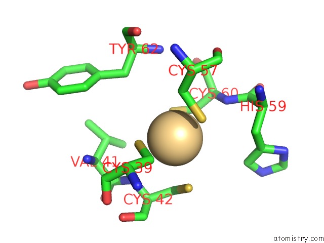
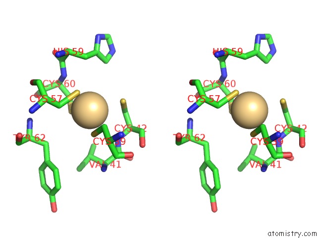
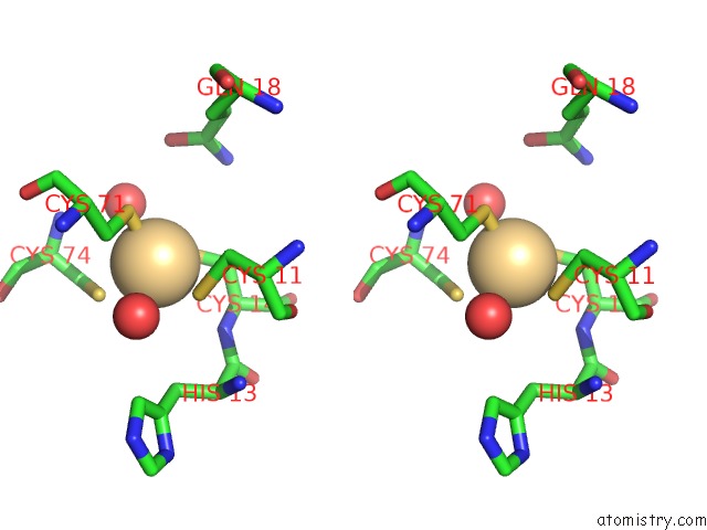
Cadmium binding site 1 out of 5 in 1q86
Go back to
Cadmium binding site 1 out
of 5 in the Crystal Structure of Cca-Phe-Cap-Biotin Bound Simultaneously at Half Occupancy to Both the A-Site and P-Site of the the 50S Ribosomal Subunit.
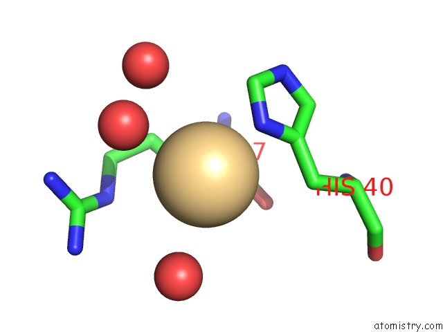
Mono view
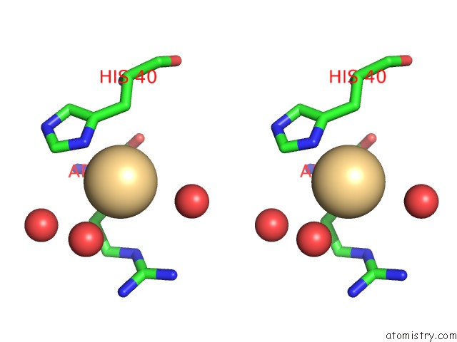
Stereo pair view
Mono view
Stereo pair view
A full contact list of Cadmium with other atoms in the Cd binding
site number 1 of Crystal Structure of Cca-Phe-Cap-Biotin Bound Simultaneously at Half Occupancy to Both the A-Site and P-Site of the the 50S Ribosomal Subunit. within 5.0Å range:
|
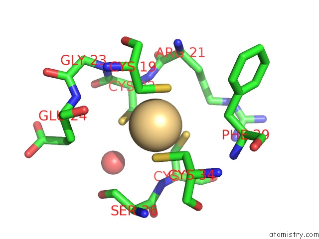
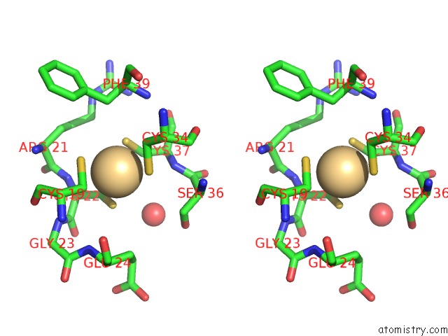
Cadmium binding site 2 out of 5 in 1q86
Go back to
Cadmium binding site 2 out
of 5 in the Crystal Structure of Cca-Phe-Cap-Biotin Bound Simultaneously at Half Occupancy to Both the A-Site and P-Site of the the 50S Ribosomal Subunit.
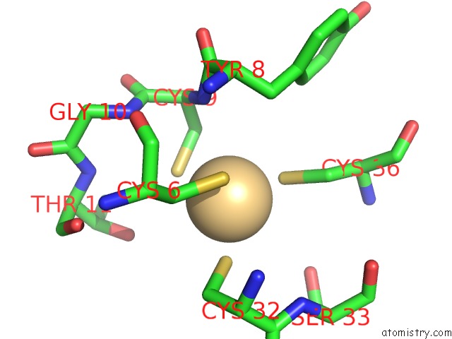
Mono view
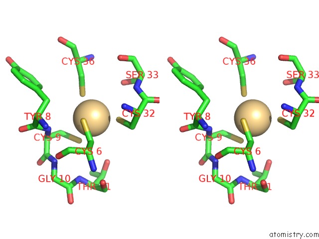
Stereo pair view
Mono view
Stereo pair view
A full contact list of Cadmium with other atoms in the Cd binding
site number 2 of Crystal Structure of Cca-Phe-Cap-Biotin Bound Simultaneously at Half Occupancy to Both the A-Site and P-Site of the the 50S Ribosomal Subunit. within 5.0Å range:
|
Cadmium binding site 3 out of 5 in 1q86
Go back to
Cadmium binding site 3 out
of 5 in the Crystal Structure of Cca-Phe-Cap-Biotin Bound Simultaneously at Half Occupancy to Both the A-Site and P-Site of the the 50S Ribosomal Subunit.
Mono view
Stereo pair view
Mono view
Stereo pair view
A full contact list of Cadmium with other atoms in the Cd binding
site number 3 of Crystal Structure of Cca-Phe-Cap-Biotin Bound Simultaneously at Half Occupancy to Both the A-Site and P-Site of the the 50S Ribosomal Subunit. within 5.0Å range:
|
Cadmium binding site 4 out of 5 in 1q86
Go back to
Cadmium binding site 4 out
of 5 in the Crystal Structure of Cca-Phe-Cap-Biotin Bound Simultaneously at Half Occupancy to Both the A-Site and P-Site of the the 50S Ribosomal Subunit.
Mono view
Stereo pair view
Mono view
Stereo pair view
A full contact list of Cadmium with other atoms in the Cd binding
site number 4 of Crystal Structure of Cca-Phe-Cap-Biotin Bound Simultaneously at Half Occupancy to Both the A-Site and P-Site of the the 50S Ribosomal Subunit. within 5.0Å range:
|
Cadmium binding site 5 out of 5 in 1q86
Go back to
Cadmium binding site 5 out
of 5 in the Crystal Structure of Cca-Phe-Cap-Biotin Bound Simultaneously at Half Occupancy to Both the A-Site and P-Site of the the 50S Ribosomal Subunit.

Mono view
Stereo pair view
Mono view
Stereo pair view
A full contact list of Cadmium with other atoms in the Cd binding
site number 5 of Crystal Structure of Cca-Phe-Cap-Biotin Bound Simultaneously at Half Occupancy to Both the A-Site and P-Site of the the 50S Ribosomal Subunit. within 5.0Å range:
|
Reference:
J.L.Hansen,
T.M.Schmeing,
P.B.Moore,
T.A.Steitz.
Structural Insights Into Peptide Bond Formation. Proc.Natl.Acad.Sci.Usa V. 99 11670 2002.
ISSN: ISSN 0027-8424
PubMed: 12185246
DOI: 10.1073/PNAS.172404099
Page generated: Thu Jul 10 11:16:15 2025
ISSN: ISSN 0027-8424
PubMed: 12185246
DOI: 10.1073/PNAS.172404099
Last articles
Cl in 3RF7Cl in 3RGF
Cl in 3RF4
Cl in 3REW
Cl in 3RE0
Cl in 3RE3
Cl in 3RDK
Cl in 3RD7
Cl in 3RDR
Cl in 3RDB